
Pathology Case of the Month - Wild Turkeys
Case History: In August 2021, a morbidity and mortality event involving both juvenile and adult Wild Turkeys (Meleagris gallopavo) was reported in Wisconsin, USA.
Sick turkeys were approachable, could be picked up without resistance, and “appeared blind”. There were no visible pox-like lesions, and birds died within hours of the initial observation. Two turkeys that were found dead were collected and submitted for necropsy examination and diagnostic testing.
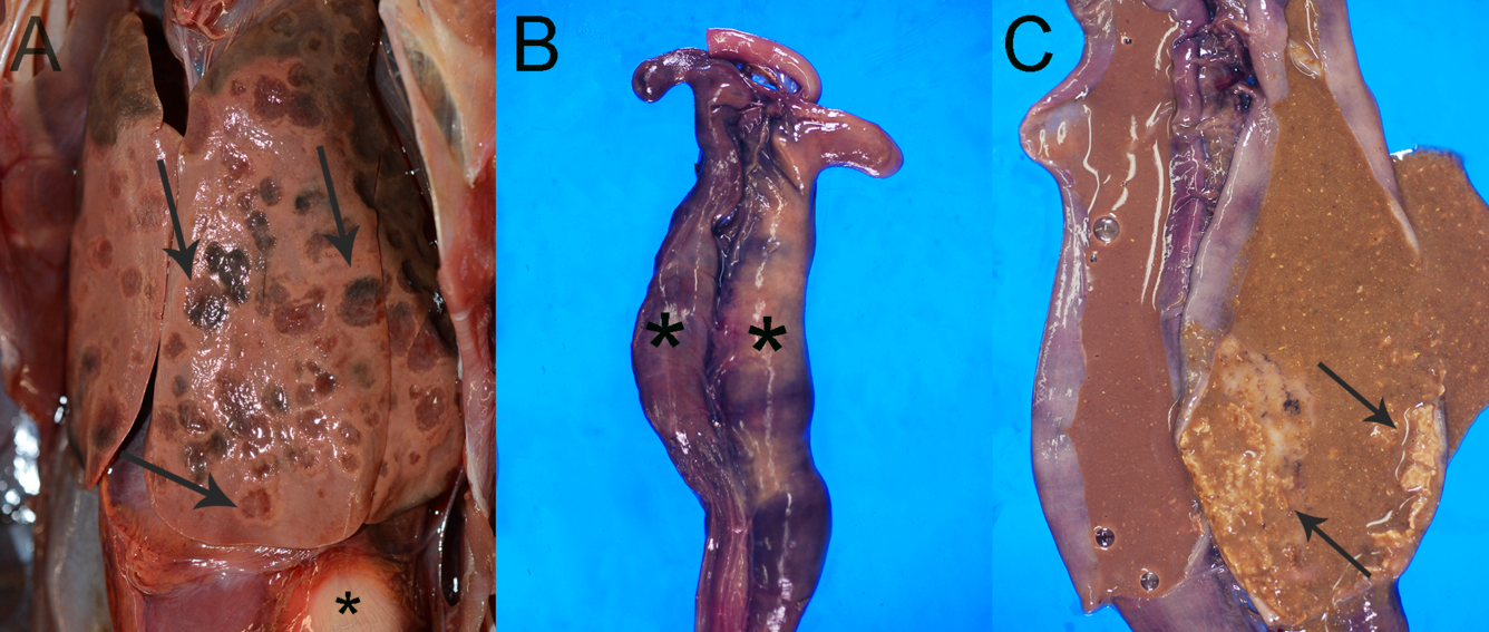
Gross Findings: Birds were in fair body condition and fair to good postmortem condition. The major gross findings in both birds involved the liver and the ceca. The liver contained multifocal to coalescing, 1-2 cm in diameter, red to brown bullseye-shaped foci (necrosis) (Fig. 1A). The paired ceca were ectatic with thickened walls, filled with tan-brown liquid content with dry, crumbly, necrotic cecal cores, and had tan plaques of necrosis on the mucosa (Fig. 1B-1C).
Histopathological Findings: Microscopic changes were similar for both birds. The liver had multifocal to coalescing foci of random hepatocellular necrosis with infiltration by macrophages, lymphocytes, plasma cells, small numbers of heterophils, and occasional multinucleated giant cells (foreign body type) (Fig. 2A). There were moderate numbers of extracellular and intrahistiocytic protozoal trophozoites (histomonads) present in the necrotic foci. Histomonads were 10-20 µm in diameter with a pale basophilic 3-5 µm diameter nucleus and often surrounded by a clear space. In the ceca, there were segmental to circumferential fibrinonecrotic membranes effacing the mucosa (Fig. 2B), and the wall was infiltrated by many macrophages, lymphocytes, and plasma cells. Histomonads, as described above, were present in the necrotic debris (Fig. 2C). In one bird, there was similar inflammation in the serosa of the proventriculus, ventriculus, ileum, and ceca, indicative of a coelomitis.

Ancillary Test(s): PCR amplification and sequencing of the partial internal transcribed spacer (ITS) 1 rRNA gene, 5.8S rRNA gene, and partial ITS2 rRNA gene was completed from swab samples collected from liver lesions from one bird and produced a 245-bp sequence that when compared to GenBank exhibited >99% identity (244/245-bp) to Histomonas meleagridis. The nematode Heterakis gallinarum was identified in the ceca from both birds based on morphology, site of infection, and sequence analysis of the 18S rRNA gene.
Morphologic Diagnoses:
- Liver: Marked multifocal to coalescing necrotizing and granulomatous hepatitis with mineralization and intrahistiocytic and extracellular protozoal trophozoites (histomonads)
- Ceca: Severe multifocal to circumferential fibrinonecrotizing and lymphohistiocytic typhlitis with extracellular and intrahistiocytic protozoal trophozoites (histomonads)
- Proventriculus, ventriculus, ileum, ceca: Moderate to marked multifocal to diffuse lymphoplasmacytic and histiocytic serositis with rare necrotic foci (1/2 birds)
Disease: Histomoniasis
Etiology: Histomonas meleagridis, a protozoan parasite
Distribution: Worldwide
Host range: Histomoniasis (black head disease) is almost exclusively a disease of Galliformes. Clinical disease is a most often reported in domestic turkeys (Meleagris gallopavo domesticus) with chickens (Gallus gallus domesticus)) often acting as asymptomatic carriers. In wild populations of birds, this disease is most commonly reported in Wild Turkeys. Domestic chickens may carry this parasite and not demonstrate any clinical signs; however, Wild Turkey, Ruffed Grouse (Bonata umbellus), Chukar (Alectoris chukar), and Peafowl (Pavo cristatus) are highly susceptible. Ring-neck Pheasant (Phasianus colchicus), Guinea Fowl (Numida meleagris), Northern Bobwhite (Colinus virginianus), Red Junglefowl (Gallus gallus), and Japanese Quail (Coturnix japonica) can also be infected. Infections in non-galliform birds are rarely reported. At the USGS National Wildlife Health Center, histomoniasis has been found in a Cackling Goose (Branta hutchinsii), Burrowing Owl (Athene cunicularia), Trumpeter Swan (Cygnus buccinator), Double-crested Cormorant (Nannopterum auritum), Tundra (Whistling) Swan (Cygnus columbianus), and Wild Turkey.
Transmission: Transmission of H. meleagridis is dependent on infection with the cecal nematode Heterakis gallinarum, which also infects many species of galliform birds. Often infection occurs from ingesting an earthworm that has ingested eggs of H. gallinarum that are in turn infected with H. meleagridis. Both parasites are released in the ceca, where H. meleagridis reproduces in the lumen. Within a week, cecal ulcerations and mixed inflammation in the ceca develop and can lead to caseous cecal cores. Histomonads enter the blood stream and are carried to the liver, where they continue to reproduce and create discrete foci of necrosis. Transmission of H. meleagridis may also occur via direct transmission or via the fecal-oral route (cloacal drinking), though the latter is mostly restricted to crowded conditions in captive populations.
Clinical signs: Birds are inactive, depressed, develop inappetence that can lead to emaciation, and stand with drooped wings, closed eyes, retracted head, and ruffled feathers. Feces may be sulfur colored or contain flecks of blood and mucus. Young birds have a more acute disease course and die within a few days after signs appear. Older birds may be sick for some time and become emaciated before death.
Pathology: Pathognomonic gross lesions involve the ceca and liver. The ceca are dilated with thickened walls, filled with yellow-green caseous exudate and/or dry, cheesy cecal cores, and have an ulcerated mucosa. Occasionally these ulcers may erode through the cecal wall and cause a coelomitis. The liver contains foci of necrosis that range from pinpoint white foci early in the disease to large bullseye-shaped foci that coalesce as the disease progresses. Microscopically, histomonads can be seen in the cecal ulcerations and foci of hepatic necrosis. Histomonads may be extracellular or within macrophages, and are round, 10-20 µm in diameter, with central dark nuclei. Histomonads can be difficult to distinguish from macrophages, and special stains such as Periodic acid-Schiff (PAS) may be needed.
Diagnosis: The gross lesions in the ceca and liver are pathognomonic. Additional testing to confirm disease can include histopathology and PCR. In this case, PCR primers specific to Histomonas were not routinely available at the USGS National Wildlife Health Center; however, the PCR method used was appropriate for the order Trichomonadida, which includes Trichomonas sp., H. meleagridis sp., Pentatrichomonas hominis, etc. Histomonads may be identified via cytology on smears of cecal contents or scrapings of cecal or liver lesions.
Public health concerns: Histomoniasis has no known public health implications.
Wildlife population impacts: The overall impact on wild galliform birds is low.
Management: There is no approved treatment or vaccine for histomoniasis. Disease management is accomplished by separating reservoir hosts (such as chickens) from vulnerable species (i.e., turkeys), and on breaking the cycle of the cecal worm vector. Caution should be used if introducing reservoir hosts into regions inhabited by vulnerable species, or if raising vulnerable species in areas where reservoir hosts have previously lived.
References:
- Beckstead RB. 2019. Histomoniasis in Poultry. Merk Veterinary Manual Online. https://www.merckvetmanual.com/poultry/histomoniasis/histomoniasis-in-poultry. Accessed October 2021.
- Crespo R, Franca MS, Fenton H, and Shivaprasad HL. 2018. Galliformes and Columbiformes. In: Pathology of Wildlife and Zoo Animals. Terio KA, McAloose D, St. Leger J, editors. Elsevier, San Diego, CA, pp. 765-767.
- Davidson WR. 2007. Histomoniasis. In: Infectious diseases of wild birds. Thomas NJ, Hunter DB, Atkinson CT, editors. Blackwell Publishing, Iowa, pp. 154-161.
- Felleisen, R.S.J. 1997. Comparative sequence analysis of 5.8S rRNA genes and internal transcribed spacer (ITS) regions of trichomonadid protozoa. Parasitology 115: 111-119. https://doi.org/10.1017/S0031182097001212
- Vieira TD, Pegoraro de Macedo MR, Bernardon FF, Müller G. 2017. Morphological, molecular and phylogenetic analyses of Diplotriaena bargusinica Skrjabin, 1917 (Nematoda: Diplotriaenidae). Parasitology International 66:555-559. https://doi.org/10.1016/j.parint.2017.04.009
Related

WHISPers

Pathology Case of the Month

Diagnostic Services
Related
WHISPers
Pathology Case of the Month