
Histopathology - Plate 6 - Figures 16-18 thumb

Detailed Description
Histopathology - Plate 6 - Figures 16-18
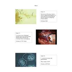
- Figure 16: Spironucleus (Hexamita) salmonis (arrow), a diplomonad flagellate, in a smear made from mucoid material from the intestine of a fingerling Chinook salmon.
- Figure 17: Ceratomyxa shasta. Perforated intestine(arrows) of a spawning fall Chinook salmon infected with Ceratomya shasta, a myxosporean. (Courtesy of J. W. Wood).
- Figure 18: C. shasta in wet mount with pansporoblasts. Phase contrast. X4500. (Courtesy of H. Wolf).