
Histopathology - Plate 9 - Figures 25-27 thumb

Detailed Description
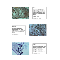
Histopathology - Plate 9 - Figures 25-27
- Figure 25: M. cerebralis infected fingerling rainbow trout with various stages of trophozoites and histo- pathological changes in the cartilagenous tissue section of the gill arch (between the arrows). (Courtesy of H. Wolf).
- Figure 26: M. cerebralis trophozoites in fingerling rainbow trout.
- Figure 27: M. cerebralis trophozoites and spores in the gill arch of fingerling rainbow trout.