
Photomicrographs from a double-crested cormorant (Nannopterum auritum) from Minnesota, USA
Detailed Description
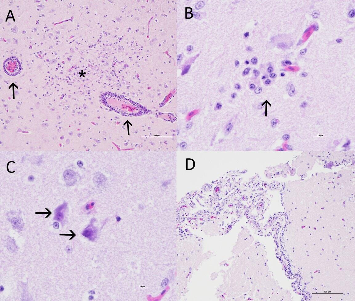
Figure 1. Photomicrographs from a double-crested cormorant (Nannopterum auritum) from Minnesota, USA. (A) Multifocal lymphocytic cuffing (arrows) and a focus of gliosis (star) within the cerebrum. H&E stain. (B) Neuronophagia in the cerebrum (arrow). H&E stain. (C) Necrotic neurons in the cerebrum (arrows). H&E stain. (D) Lymphocytic meningitis in the cerebellum. H&E stain.
Sources/Usage
Public Domain.
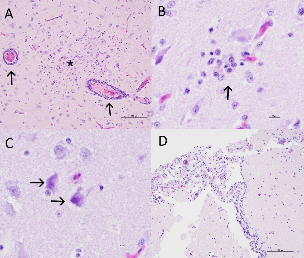
Figure 1. Photomicrographs from a double-crested cormorant (Nannopterum auritum) from Minnesota, USA. (A) Multifocal lymphocytic cuffing (arrows) and a focus of gliosis (star) within the cerebrum. H&E stain. (B) Neuronophagia in the cerebrum (arrow). H&E stain. (C) Necrotic neurons in the cerebrum (arrows). H&E stain. (D) Lymphocytic meningitis in the cerebellum. H&E stain.