
Amphimerus elongatus in a Bald Eagle (Haliaeetus leucocephalus)
History: An immature 2860-g Bald Eagle (Haliaeetus leucocephalus) from South Carolina, USA was found weak and lethargic with labored breathing, a head tilt and nystagmus and was taken to a rehabilitation facility. The bird died hours after admission and was submitted for necropsy.
Gross Findings: On external examination, a 1 x 0.5 cm wound was present in the skin over the right caudal ribs; it did not extend into the coelomic cavity. Lice were present in feathers of the head. Green fecal material was present in feathers around the vent. On internal examination, there was minimal subcutaneous, no visceral and a small amount of epicardial fat. Pectoral muscle was severely atrophied. The esophagus, proventriculus and ventriculus and intestines contained brown gritty blood. The liver was mottled red and light brown with numerous dark green serpentine foci (Fig. 1 A,B). Kidneys were light brown-green with visible urates on cut section.
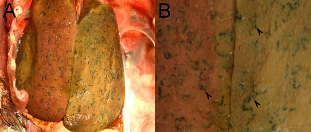
Figure 1. Photographs from a Bald Eagle (Haliaeetus leucocephalus) from South Carolina, US. (A) Numerous green serpentine foci are present throughout the liver. (B) Higher magnification of the liver showing green serpentine foci (arrowheads).
Histopathological Findings: Approximately 50% of the hepatic parenchyma is replaced by large trematodes laden with numerous eggs (Fig. 2A,B). In some foci, collections of eggs, occasionally admixed with orange pigment and bacteria, are surrounded by necrotic cellular debris rimmed by macrophages and further surrounded by multinucleated giant cells (Fig 2C). Accumulations of fluke pigment are also observed (Fig. 2D). Scattered throughout the liver are foci of eosinophilic debris admixed with bacteria surrounded by multinucleated giant cells. Collections of trematode eggs, as observed in the liver, are scattered throughout the pancreas along with foci of necrosis and inflammation.
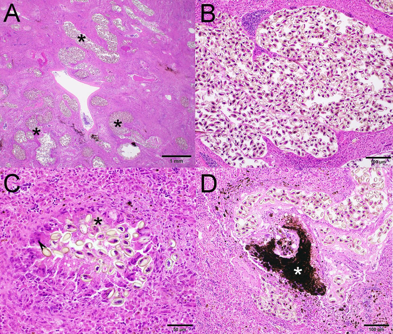
Figure 2. Photomicrographs from a Bald Eagle (Haliaeetus leucocephalus) from South Carolina, USA. H&E stain. (A) Low magnification of the liver shows approximately 50% of the hepatic parenchyma is replaced by large trematodes laden with numerous eggs (asterisks). (B) Higher magnification showing trematodes eggs. (C) A cluster of trematode eggs surrounded by bacteria (asterisk), epithelioid macrophages and multinucleated giant cells (arrowhead). (D) Fluke pigment (asterisk) is occasionally observed in the liver.
Etiology: Amphimerus elongatus, a digenetic trematode of the Opisthorchiidae family.
Host range: Found in Double-crested Cormorants (Nannopterum auritum), Common Loons (Gavia immer), Bald Eagles, Belted Kingfishers (Megaceryle alcyon), Whooping Cranes (Grus americana), ducks and swans.
Life cycle: Digenean trematodes have complex life cycles, often using a mollusk as the first intermediate host, other invertebrates and vertebrates as a second intermediate host, whereas the definitive hosts are often vertebrates, such as various fishes, amphibians, reptiles, birds and mammals. The sexual phase occurs in the vertebrate definitive host and the asexual phase in the molluscan intermediate host.
Transmission: Eggs are produced in the definitive host and are passed in feces→a miracidium exits the egg and infects an intermediate host (usually a snail)→the miracidium develops into a sporocyst which reproduces asexually to form more sporocysts or rediae→rediae reproduce asexually to form more rediae or cercariae→cercariae emerge from the snail and penetrate a second intermediate host, the definitive host or encyst on vegetation where they form metacercariae→adult worms develop after the definitive host ingests metacercariae.
Geographic distribution: The trematode is found anywhere the intermediate hosts occur.
Pathology: Gross lesions include hepatomegaly with irregular capsular surfaces and numerous white, dark green, or black foci and tracts. Microscopic lesions include granulomas with intralesional necrotic debris, trematode eggs, trematode pigment, and, occasionally, bacterial colonies in the liver or pancreas. Gravid trematodes can compress hepatocytes in portal areas.
Diagnosis: Histopathology; dissection scope examination; PCR.
Public health concerns: Liver flukes of the genus Amphimerus have been shown to infect humans, suggesting these trematodes are zoonotic pathogens.
Wildlife population impacts: While infections can be fatal in individual birds, there are not likely significant impacts on wildlife populations.
Management: Control measures can involve reduction of the snail intermediate host.
References:
Boyd EM & Fry AE. 1971. Metazoan parasites of the Eastern belted kingfisher, Megaceryle alcyon alcyon. J Parasitol 57 (1): 150–156. DOI: 10.2307/3277771.
Calvopiña M, Cevallos W, Kumazawa H & Eisenberg J. 2011. High prevalence of human liver infection by Amphimerus spp. flukes, Ecuador. Emerg Infect Dis 17(12):2331–2334. DOI: 10.3201/eid1712.110373.
Huffman JE. 2008. Trematodes. In: Parasitic Diseases of Wild Birds, Atkinson CT, Thomas NJ, Hunter DB, editors. Wiley-Blackwell, Ames, Iowa, pp 225–245.
Pense DB & Childs GE. 1972. Pathology of Amphimerus elongatus (Digenea: opisthorchiidae) in the liver of the double-crested cormorant. J Wild Dis 8: 221–224.
Related

WHISPers

Pathology Case of the Month

Diagnostic Services
Related
WHISPers
Pathology Case of the Month