



Heterotopic mesenteric ossification in a Northern Sea Otter (Enhydra lutris kenyoni)
History: An aged adult male 36.8 kg Northern Sea Otter (Enhydra lutris kenyoni) from Washington, USA was found dragging its hind end. It was humanely euthanized and the carcass was submitted for necropsy.
Gross Findings: There were no significant external findings. On internal examination, there was no subcutaneous, visceral, peri-renal and epicardial fat. The heart was enlarged and small smooth nodules were present on the left atrioventricular (AV) valve. The liver was enlarged with a nutmeg pattern on cut surface. Three irregular tan pieces of hard bone were present in the mesentery around the mesenteric lymph node (Fig. 1 A, B).
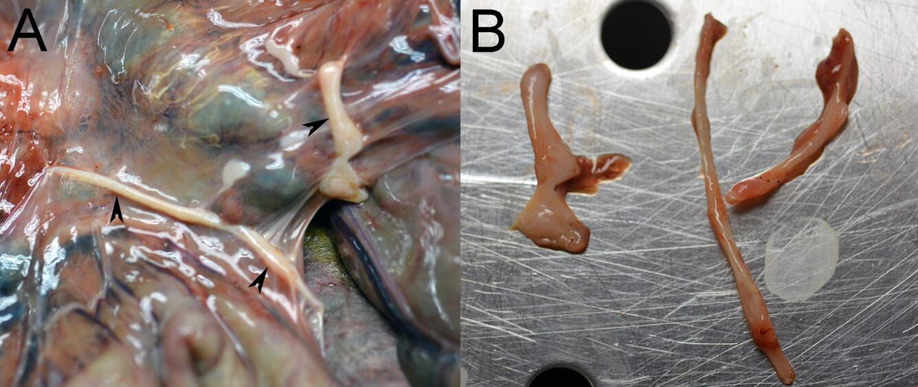
Figure 1. Photographs from a Northern Sea Otter (Enhydra lutris kenyoni) from Washington, USA. (A) Irregular, bony structures (arrowheads) are present in the mesentery surrounding the mesenteric lymph node. (B) Three irregular bony structures are removed from the mesentery.
Diagnosis: The cause of morbidity for which euthanasia was performed in this sea otter was heart failure. Heterotopic mesenteric ossification was diagnosed incidentally. Heterotopic mesenteric ossification is a benign growth of bone that occurs in the mesentery. In heterotopic ossification, osteoblasts form lamellar bone at an extraskeletal site through the deposition, remodeling and mineralization of osteoid.
Cause: In humans, severe repetitive abdominal injuries or abdominal surgery are thought to lead to reactive bone formation in the mesentery.
Host range: In humans, cases occur in older adults and more commonly in males. A case in a sow led to death after mesenteric torsion around the heterotopic bone. Another report described an intestinal volvulus in a sow associated with heterotopic mesenteric ossification. A ferret that presented with anorexia, weakness and weight loss had firm, palpable abdominal masses that were identified as bone after surgical removal.
Pathology: Gross lesions include hard bony tissues with sharp edges in the intestinal mesentery. Microscopically, the structures are composed of trabecular bone.
Pathogenesis: While the pathogenesis is not known, in humans it is thought that four criteria are necessary to allow heterotopic mesenteric ossification to occur: (1) a triggering event; (2) a signal released by the trigger; (3) an energy supply and signal to proliferating mesenchymal cells to transform into chondroblasts and osteoblasts; and (4) a suitable environment. In humans, it is thought that trauma, ischemia, or intraabdominal infection causes osteoprogenitor cells to differentiate into osteoblasts and induce bone formation. It is also theorized that it may occur when bone periosteum implants into soft tissue and proliferates to form bone.
Diagnosis: Gross examination; histopathology.
Public health concerns: Heterotopic mesenteric ossification can cause bowel obstruction and mortality in rare cases. Cases are rare with only 73 reported in the human literature from 1983 to 2020.
Wildlife population impacts: There are not likely significant impacts on wildlife populations.
References:
Althaqafi RMM, Assiri SA, Aloufi RA, Althobaiti F, Althobaiti B, & Al AdwaniM. 2021. A case report and literature review of heterotopic mesenteric ossification. Int J Surg Case Rep, 82: 105905. DOI:10.1016/j.ijscr.2021.105905.
Corbetta D, Degl'Innocenti S, Bianco C, Iveson T, Ortiz A, Anselmi C, Morey-Matamalas A. 2024. Intra-abdominal heterotopic mesenteric ossification associated with intestinal volvulus in a sow (Sus scrofa domesticus). J Comp Pathol, 215:32–36. DOI:10.1016/j.jcpa.2024.09.003.
Lei J, McCoy CS, Luo H, Harrington Jr EP, Rosales I, Poznansky MC, Markmann JF. 2017. Heterotopic ossification of laparotomy scars in nonhuman primate. J Surg, 2017(02):1–4. DOI: 10.29011/JSUR-117.000017.
Miller MA & Zachary JF. 2017. Mechanisms and morphology of cellular injury, adaptation, and death. In: Pathologic Basis of Veterinary Disease, 6th ed, Zachary JF, editor. Elsevier, St. Louis, Missouri, p 35.
Sanford SE & Rehmtulla AJ. 1994. Heterotopic mesenteric bone: An unexpected cause of mesenteric torsion in a sow. J Swine Health Prod, 2(1):17–18.
Tecilla M, Bielli M, Savarino P, & Roccabianca P. 2018. Mesenteric heterotopic ossificans in a ferret (Mustela putorius furo): A rare cause of soft tissue ossification. J Exoti Pet Med, 27:108–112. DOI:10.1053/j.jepm.2018.02.003.
Related

WHISPers

Pathology Case of the Month

Diagnostic Services
Related
WHISPers
Pathology Case of the Month