
Renal Coccidiosis in Snow Geese from California, USA
Author(s): Marcos Isidoro-Ayza
History: Two juvenile snow geese (Anser caerulescens) were found dead at a National Wildlife Refuge (NWR) in California, US. The site consists of seasonal wetland habitat for waterbirds. The birds had been observed on the refuge and at neighboring hunt clubs with clinical signs including spinning behavior, droopy heads, and separation from flocks.
Gross Findings: Both birds were in poor postmortem condition with moderate to advanced autolysis. External examination revealed no significant abnormalities. Internally, both had a small amount of subcutaneous and visceral fat, indicating fair body condition. The kidneys were bilaterally enlarged, pale, and mottled, with multifocal to coalescing pinpoint white foci throughout the parenchyma.
Histopathological Findings: In the renal tissue examined, >75% of the area showed degeneration and necrosis of renal tubular epithelium, particularly affecting the collecting ducts. Tubular epithelial cells exhibited swelling, karyolysis, and sloughing into the lumen. Tubules were frequently distended by necrotic cellular debris, uric acid crystals, and abundant coccidia developmental stages compatible with Eimeria sp. (Fig. 1A). Numerous intracellular and intraluminal parasitic forms were present, including macrogametocytes, microgametocytes, and oocysts. These organisms caused marked expansion and distortion of tubular epithelial cells, often resulting in cell rupture (Fig. 1B).
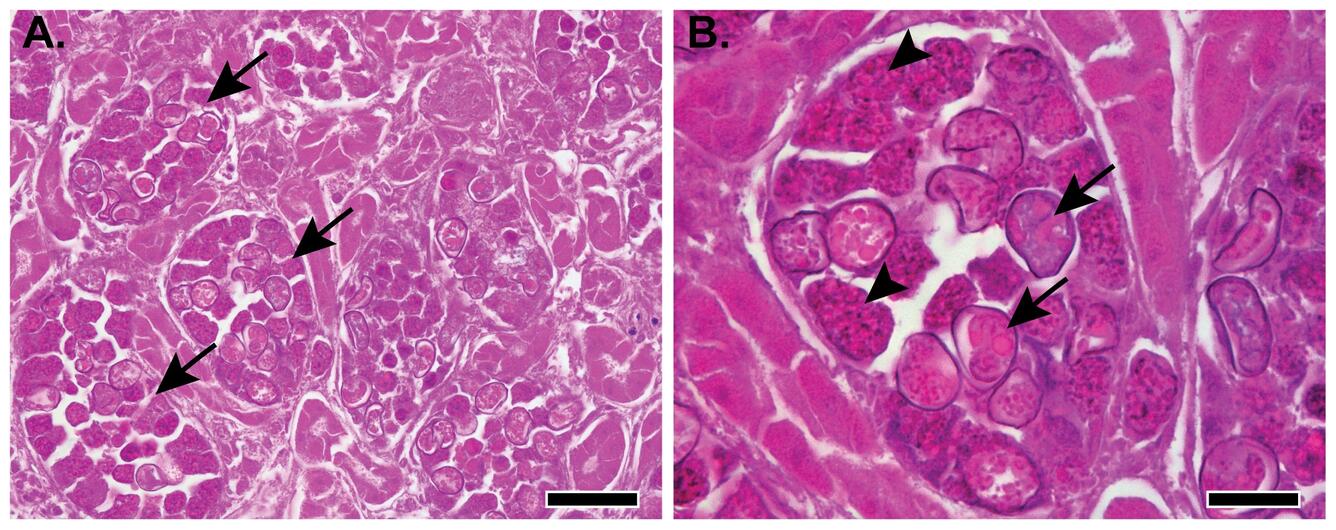
Figure 1. Photomicrographs from the kidney of one of the two snow geese (Anser caerulescens) found dead in California, US. (A) Multifocal to coalescing degeneration and necrosis of over 75% of the renal tubular epithelium with intralesional coccidia compatible with Eimeria sp. (arrows). Autolytic changes are present but do not obscure the primary pathological lesions. H&E stain. Scale bar=100µm. (B) Intracellular and intraluminal parasitic forms, including macrogametocytes (arrow heads), and oocysts (arrows). H&E stain. Scale bar=20µm.
Morphologic Diagnosis: Necrotizing tubular nephritis with abundant intralesional coccidia (Eimeria sp.), subacute, generalized, severe.
Cause of death: Acute renal failure secondary to severe renal coccidiosis causing extensive tubular damage and loss of functional renal parenchyma. The neurological signs described in the history, including spinning behavior and droopy heads were likely secondary to toxemia, dehydration, or electrolyte disturbances associated with severe renal dysfunction.
Disease: Renal coccidiosis
Etiology: Protozoal parasites of the genus Eimeria, which infects renal tubular epithelium and the ureters. In snow geese, the disease is most often associated with Eimeria truncata.
Distribution: Worldwide.
Seasonality: Occurs any time of year. Mortality is most common when birds are densely aggregated, such as on breeding or wintering grounds.
Host range: Primarily waterfowl, including snow geese and other geese and ducks.
Other bird species may also be affected depending on the Eimeria species.
Life cycle: Renal Eimeria spp. have a direct life cycle. Infected birds shed oocysts in urine and feces. Within 1–5 days, the sporont divides to form four sporocysts, each containing two sporozoites (eight total). A bird ingests sporulated oocysts; sporozoites are released in the digestive tract, penetrate the intestinal wall, enter the bloodstream, and travel to the kidneys. There, they invade renal tubular epithelium and undergo schizogony and gametogony, producing unsporulated oocysts that are shed in urine and feces, completing the cycle.
Transmission: Birds become infected through ingestion of sporulated oocysts in contaminated water, soil, or food. High-density aggregations promote fecal-urine contamination of the environment, increasing exposure risk.
Clinical signs: Variable and nonspecific. Severe cases may show weakness, lethargy, anorexia, emaciation, and, less commonly, neurologic signs secondary to renal failure, particularly in juveniles or stressed individuals.
Pathology:
Gross Lesions: Kidneys are enlarged, pale, and mottled, with multifocal white foci throughout the renal parenchyma. The cut surface may reveal chalky urate deposits.
Microscopic Lesions: Protozoal developmental stages (schizonts, gametocytes, and oocysts) are present within renal tubules and ureters, accompanied by tubular necrosis, and uric acid crystal accumulation. Most cases of renal coccidiosis are accompanied by a mild to moderate lymphoplasmacytic interstitial nephritis, but interstitial inflammation was minimal to absent in this case, likely reflecting the acute/subacute course and dominance of extensive tubular necrosis.
Diagnosis:
Suspected diagnosis. Based on compatible gross lesions (pale, mottled, enlarged kidneys), history of affected juveniles in dense populations.
Definitive diagnosis. Requires microscopic lesions and identification of Eimeria sp. stages in kidney/ureter.
Autolysis interference: These snow geese were recovered in poor postmortem condition, with moderate to advanced autolysis. This is common in wildlife pathology where time of death is unknown, and carcasses are exposed to environmental conditions before retrieval. Despite this, renal lesions were severe and characteristic, allowing a definitive diagnosis. Autolytic changes were present but did not obscure the primary lesions or parasitic structures.
Public health concerns: No known human health risk.
Wildlife population impacts: Severe renal coccidiosis can result in significant mortality in juvenile birds and, less commonly, in stressed or immunocompromised adults, particularly in high-density aggregations where environmental contamination is amplified. Renal coccidiosis has previously been described as an incidental finding in adult geese, including a prior Case of the Month (Renal Coccidiasis in a Canada Goose [Branta canadensis] from Ohio, USA). In contrast, the present case demonstrates the potential for fatal disease in juveniles. The extent of renal injury observed in this case is expected to result in severe renal dysfunction and acute renal failure. Immature geese are immunologically naïve and less capable of limiting parasite replication, leading to higher parasite burdens, extensive tubular damage, and an increased risk of mortality.
Management: 1) Reducing crowding and improving habitat quality to decrease contamination. 2) Carcass removal in high-density areas (if feasible). 3) Control in wild populations is generally not feasible
References:
United States Geological Survey. 1999. Field manual of wildlife diseases: general field procedures and diseases of birds, https://pubs.er.usgs.gov/publication/itr19990001. Accessed April 2025.
Yabsley MJ. 2008. Eimeria. In: Parasitic Diseases of Wild Birds, Atkinson CT, Thomas NJ, Hunter DB, editors. Wiley-Blackwell, Ames, Iowa, pp 162–180.
Related

WHISPers

Pathology Case of the Month

Diagnostic Services
Related
WHISPers
Pathology Case of the Month