
Expanded vermiculite ores examined in this study

Detailed Description
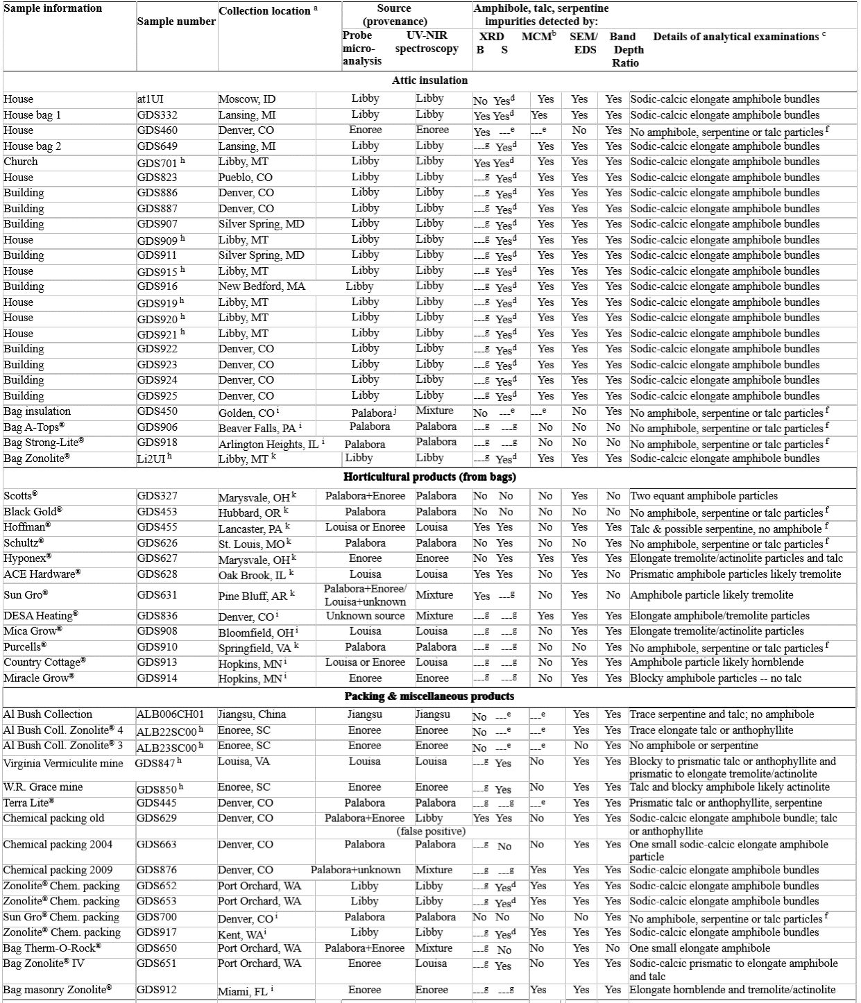
Table 1. Expanded vermiculite ores examined in this study: their sources and amphibole, talc, and/or serpentine impurities.
Notes: XRD = X-ray diffraction analysis of bulk ore samples (B) and sink fractions (S). SEM-EDS = Scanning Electron Microscope – Energy-Dispersive X-Ray Spectroscopy. Band Depth Ratio = 1.40/1.42-µm band depth ratio. The use of "sodic-calcic" refers to amphiboles with compositions including primarily those of winchite, rictherite, and tremolite. The exact amphibole species could not be determined with qualitative SEM-EDS analysis (Meeker et al. 2003; Lowers and Meeker 2004). In this study, we use the term “elongate amphibole particle” (NIOSH 2011) to encompass all amphibole crystals longer than 5 µm with an aspect ratio greater than 3:1 whose habits are asbestiform or nonasbestiform or may have formed as a result of cleaving. Unknown source = distinctly different spectral signature than any of those of the four major historical sources of expanded vermiculite ore. Mixture = combination of ores, one of which is from one of four major historical sources of commercial expanded vermiculite ore. Enoree = Enoree, South Carolina; Libby = Libby, Montana; Louisa = Louisa, Virginia; Palabora = Palabora Mine in South Africa. Chem. = chemical; comp. = composition.
a Sample collection location is given; otherwise city of purchase i or product manufacture k is provided when available.
b MCM = Modified Cincinnati Method (see text for description). Yes = detection of macroscopically visible elongate amphibole, talc, or serpentine.
c Details of MCM, microprobe, spectroscopy, XRD, and SEM-EDS analyses for expanded vermiculite ore samples can be found in the individual sample descriptions in Swayze et al. (2018).
d Macroscopic elongate amphibole bundles were removed from the sink fraction prior to XRD analysis but were verified as amphibole by SEM analysis, thus they are listed here as being “detectable” by XRD.
e Insufficient sample volume to generate a sink fraction for analysis with XRD or MCM.
f If hours of searching revealed no amphibole, talc, and/or serpentine particles on an SEM mount then the sample was not considered to contain them.
g XRD analysis not performed on the bulk sample and/or its sink fraction sample as noted.
h Samples, among others (Lowers and Meeker 2004), used to establish provenance fields on electron probe microanalysis diagrams (Fig. 4).
i See a above.
j Large Palabora flakes spatially dominate probe mounts so analyses may not have sampled smaller flakes from a non-Palabora source in this mixture of ores.
k See a above.
Sources/Usage
Public Domain.
Characterizing the source of potentially asbestos-bearing commercial vermiculite insulation using in situ IR spectroscopy
Swayze, G.A., Lowers, H.A., Benzel, W.M., Clark, R.N., Driscoll, R.L., Perlman, Z.S., Hoefen, T.M., and Dyar, M.D., 2018, American Mineralogist, v. 103, p. 517-549.
https://doi.org/10.2138/am-2018-6022
Abstract:
Commercially produced vermiculite insulation from Libby, Montana, contains trace
levels of asbestiform amphibole, which is known to cause asbestos-related diseases. When
vermiculite insulation is found in a building, evaluation for its potential asbestos content
traditionally involves collecting a sample from an attic or wall and submitting it for potentially
time-consuming analyses at an off-site laboratory. The goal of this study was to determine if in
situ near-infrared reflectance measurements could be used to reliably identify the source of
vermiculite ore and therefore its potential to contain asbestos. Spectra of 52 expanded ore
samples, including attic insulation, commercial packing materials, and horticultural products
from Libby, Montana; Louisa, Virginia; Enoree, South Carolina; Palabora, South Africa; and
Jiangsu, China, were measured with a portable spectrometer. The mine sources for these
vermiculite ores were identified based on collection location, when known, and on differences in
elemental composition as measured by electron probe microanalysis. Reflectance spectra of the
insulation samples show vibrational overtone and combination absorptions that vary in
wavelength position and relative intensity depending on elemental composition and proportions
of their constituent micas (i.e., vermiculite ore usually consists of a mixture of hydrobiotite and
vermiculite mineral flakes). Band depth ratios of the 1.38/2.32-, 1.40/1.42-, and 2.24/2.38-µm
absorptions allow determination of a vermiculite insulation’s source and detection of its potential
to contain amphibole, talc, and/or serpentine impurities. Spectroscopy cannot distinguish
asbestiform vs non-asbestiform amphiboles. However, if the spectrally determined mica
composition and mineralogy of an insulation sample is consistent with ore from Libby, then it is
likely that some portion of the sodic-calcic amphibole it contains is asbestiform, given that all of
the nearly two dozen Libby vermiculite insulation samples examined with scanning electron
microscopy in this study contain amphiboles. One sample of expanded vermiculite ore from
multiple sources was recognized as a limitation of the spectral method, therefore an additional
test (i.e., 2.24-µm absorption position vs 2.24/2.38-µm band depth) was incorporated into the
spectral method to eliminate misclassification caused by such mixtures. With portable field
spectrometers, the methodology developed can be used to determine vermiculite insulation’s
source and estimate its potential amphibole content, thereby providing low-cost analysis with
onsite reporting to property owners.