



fibrinoid vascular necrosis_Photomicrographs from a Barred Tiger Salamander (Ambystoma mavortium) found dead in Idaho, USA.
Detailed Description
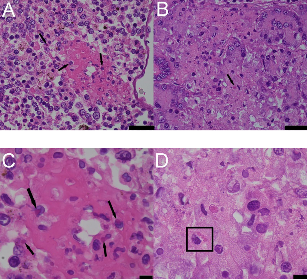
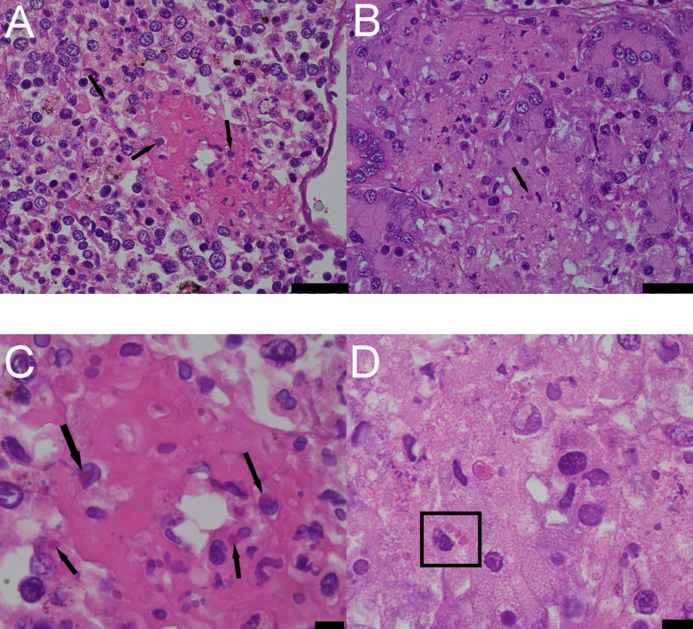
Figure 2. Photomicrographs from a Barred Tiger Salamander (Ambystoma mavortium) found dead in Idaho, USA. A. Focused on an area of fibrinoid vascular necrosis in the spleen, there are numerous eosinophilic intracytoplasmic inclusion bodies (black arrows) with cellular necrosis. H&E stain. Scale bar = 40µm. B. Focused on an area of tubular necrosis in the kidney, there are numerous eosinophilic intracytoplasmic inclusion bodies. H&E stain. Scale bar = 40µm. C and D. 100x view of the intracytoplasmic inclusion bodies in the spleen (black arrows) and kidney (square). H&E. Scale bars = 10 µm.
Sources/Usage
Public Domain.
Figure 2. Photomicrographs from a Barred Tiger Salamander (Ambystoma mavortium) found dead in Idaho, USA. A. Focused on an area of fibrinoid vascular necrosis in the spleen, there are numerous eosinophilic intracytoplasmic inclusion bodies (black arrows) with cellular necrosis. H&E stain. Scale bar = 40µm. B. Focused on an area of tubular necrosis in the kidney, there are numerous eosinophilic intracytoplasmic inclusion bodies. H&E stain. Scale bar = 40µm. C and D. 100x view of the intracytoplasmic inclusion bodies in the spleen (black arrows) and kidney (square). H&E. Scale bars = 10 µm.