



Ranavirus in a Barred Tiger Salamander (Ambystoma mavortium)
History: Mass mortality reported in Barred Tiger Salamanders (Ambystoma mavortium) in Idaho, USA. Clinical signs include ataxia, erratic swimming, inability to stay upright, and lethargy.
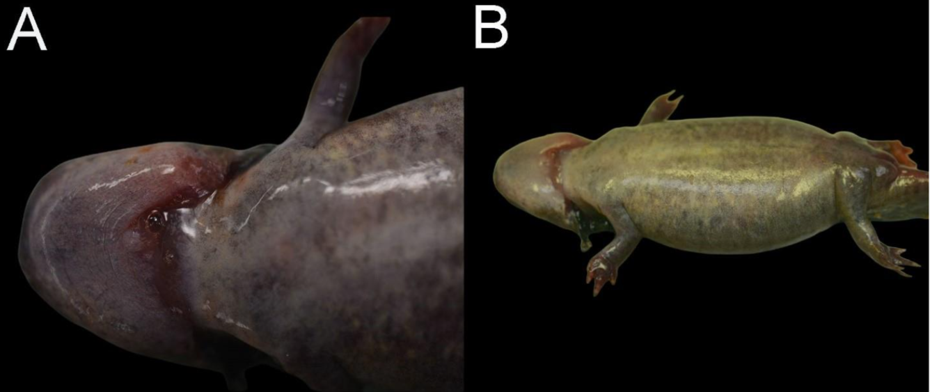
Gross findings: The carcass was submitted fixed in 10% neutral buffered formalin without prior external examination. One salamander that was submitted fresh for necropsy had sloughing skin around the feet, reddening of the gular and vent regions, and swollen abdomen.
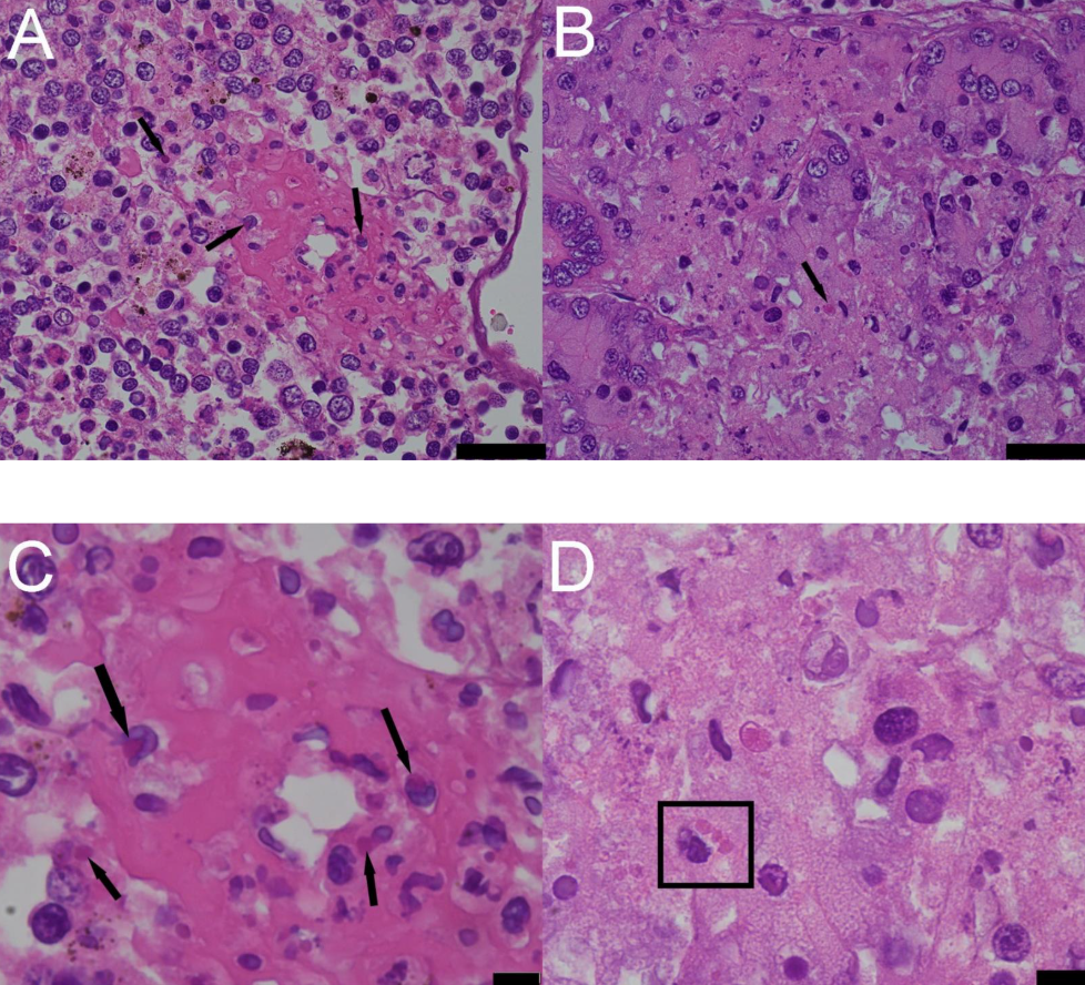
Histopathological findings: There are multifocal to coalescing, randomly distributed foci of necrosis in the spleen, kidney, skin, oral mucosa, and intestinal epithelium. Multifocal fibrinoid necrosis of small vessels was observed, characterized by eosionophilic deposition within the vessel wall and associated with inflammatory infiltrates. Intracytoplasmic inclusion bodies are present in the spleen, kidney, liver and skin.
Morphologic diagnoses: Necrotizing dermatitis, splenitis, nephritis, acute, multifocal to coalescing, random, severe, with intracytoplasmic eosinophilic inclusion bodies (consistent with Ranavirus).
Disease: Ranavirosis
Etiology: Ranavirus (most likely Ambystoma tigrinum virus (ATV)), Iridoviridae; DDx: Other ranaviruses (Frog Virus 3 (FV3), Bohle iridovirus (BIV), Midwife toad virus).
Seasonality: Late summer to early autumn (for ATV).
Distribution and Host Range:
ATV: North America; primarily affects Caudata of all life stages.
FV3: Worldwide; affects amphibians primarily, reptiles, and fish.
BIV: Affects fish primarily and amphibians.
Midwife toad virus: Affects amphibians.
Transmission: Direct contact between animals, contaminated water, and ingestion of infected carcasses, potential mosquito transmission of FV3 to terrestrial turtles.
Clinical Signs: Sudden death or signs such as ataxia, lethargy, erratic swimming, inability to maintain upright posture.
Pathology:
Gross: Skin erythema, ulcers, subcutaneous edema, petechia and ecchymoses of the skin and viscera and ascites. Raised cutaneous plaques may be often associated and present with ATV.
Histologically: Necrosis and vasculitis of hematopoietic tissue and visceral organs and intracytoplasmic eosinophilic inclusion bodies.
Diagnosis:
Suspect: Gross and histologic compatible lesions with eosinophilic intracytoplasmic inclusion bodies in affected tissues.
Confirmed: Gross and histologically compatible lesions and direct viral detection by PCR, immunohistochemistry, in situ hybridization, or viral isolation from tissues followed by PCR or sequencing.
Public Health Concerns: None; currently, there are no known direct risks to humans.
Wildlife population impacts: Ranavirus has caused mass mortality of amphibians since the early 1990s, affecting a wide range of species resulting in 90 to 100% mortality in any given outbreak. This has had a massive impact on endangered amphibian species across the world in wild and captive populations that the virus has now been listed as notifiable disease for the World Organization of Animal Health (WOAH) in amphibians and fish. Due to causing massive population declines, this virus has resulted in strong selective pressure in their hosts, which may have impacts to come if populations cannot develop immunity.
Management: Restrict movement of amphibians from different locations and decontamination of field equipment for all studies involving aquatic environments with amphibians present and avoid uses in multiple locations.
References:
Garcês A, Pires I. 2024. Ranavirus and Its Impact on the Amphibian Population. Middle East Research Journal of Biological Sciences 4:127-138. doi: 10.36348/merjbs.2024.v04i04.005.
Marschange RE. 2019. Virology. In: Mader’s Reptile and Amphibian Medicine 3rd ed. Divers SJ, Stahl SJ, editors. Elsevier, Missouri, pp. 247-269.
Miller D, Gray M, Storfer A. 2011. Ecopathology of ranaviruses infecting amphibians. Viruses 3(11): 2351-2373. doi: 10.3390/v3112351.
OIE. 2016. Aquatic Animal Health Code [WWW Document] (Accessed 10 March 2026)..
Oura C. 2017. Asfarviridae and Iridoviridae. In: Fenner’s Veterinary Virology 5th ed. MachLachlan NJ, Dubovi EF, editors. Academic Press, Massachusettes, pp. 175-188.
Pessier AP. 2018. Amphibia. In: Pathology of Zoo and Wildlife. Terio KA, McAloose D, St. Leger J, editors. Academic Press, Massachusetts, pp. 915-944.
Price SJ, et al. 2017. From fish to frogs and beyond: Impact and host range of emergent ranaviruses, Virology 511: 272-279.
Related

WHISPers

Pathology Case of the Month

Diagnostic Services
Related
WHISPers
Pathology Case of the Month