
Figure 2. Photomicrographs from a raccoon (Procyon lotor) found in Wisconsin, USA.
Detailed Description
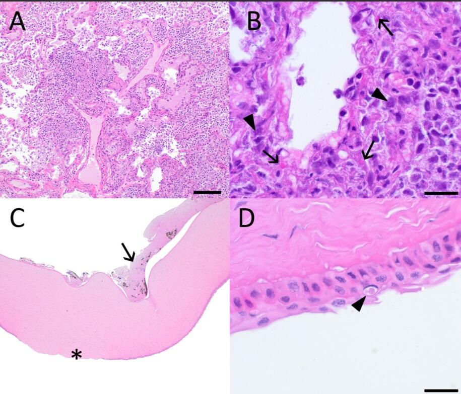
Figure 2. Photomicrographs from a raccoon (Procyon lotor) found in Wisconsin, USA. (A) Lung. Alveoli are filled with and sometimes effaced by numerous degenerate and nondegenerate neutrophils, macrophages, fibrin, proteinaceous fluid, and necrotic debris. Septa are expanded by fibrin and inflammatory cells. Bar = 100 µm. (B) High magnification of 2A showing macrophages and fewer degenerate and nondegenerate neutrophils, necrotic debris, and intranuclear (arrowheads) and intracytoplasmic (arrows) amorphous eosinophilic viral inclusions within macrophages and epithelial cells. Bar = 20 µm. (C) Cornea. Corneal epithelium is ulcerated and there is edema of the corneal stroma (asterisk). Fibrin containing melanin that is free and within neutrophils is adhered along the endothelial border. (D) High magnification of corneal epithelium. Squamous epithelium is eroded, and a large, amorphous eosinophilic cytoplasmic inclusion is present (arrowhead). Bar = 20 µm.
Sources/Usage
Public Domain.
Figure 2. Photomicrographs from a raccoon (Procyon lotor) found in Wisconsin, USA. (A) Lung. Alveoli are filled with and sometimes effaced by numerous degenerate and nondegenerate neutrophils, macrophages, fibrin, proteinaceous fluid, and necrotic debris. Septa are expanded by fibrin and inflammatory cells. Bar = 100 µm. (B) High magnification of 2A showing macrophages and fewer degenerate and nondegenerate neutrophils, necrotic debris, and intranuclear (arrowheads) and intracytoplasmic (arrows) amorphous eosinophilic viral inclusions within macrophages and epithelial cells. Bar = 20 µm. (C) Cornea. Corneal epithelium is ulcerated and there is edema of the corneal stroma (asterisk). Fibrin containing melanin that is free and within neutrophils is adhered along the endothelial border. (D) High magnification of corneal epithelium. Squamous epithelium is eroded, and a large, amorphous eosinophilic cytoplasmic inclusion is present (arrowhead). Bar = 20 µm.