
Photomicrographs from a Canada Goose (Branta canadensis) from Ohio, USA.
Detailed Description
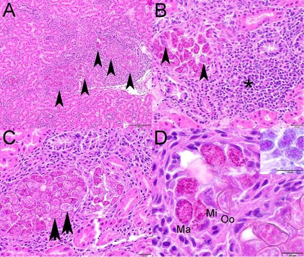
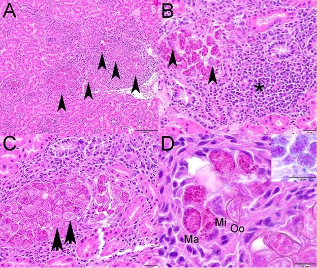
Photomicrographs from a Canada Goose (Branta canadensis) from Ohio, USA. Hematoxylin and eosin stain. (A) Renal tubules are distended with various stages of intraepithelial coccidia (arrowheads) and are surrounded by inflammation. (B) Macrogamonts and fewer microgamonts (arrowheads) expand the cytoplasm of renal tubular epithelial cells and the affected tubule is surrounded by large numbers of lymphocytes and plasma cells and fewer macrophages and granulocytes (asterisk). (C) Large numbers of coccidial oocysts (arrowheads) fill the cytoplasm of renal tubular epithelial cells. (D) Macrogamonts (Ma) with peripheral PAS positive granules (inset), microgamonts (Mi), and unsporulated oocysts with a thick wall (Oo) are present in the cytoplasm of renal tubular epithelial cells.
Sources/Usage
Public Domain.
Photomicrographs from a Canada Goose (Branta canadensis) from Ohio, USA.