
Renal Coccidiasis in a Canada Goose (Branta canadensis) from Ohio, USA
History: An immature male 2460-g Canada Goose (Branta canadensis) was euthanized as part of a mortality event in Ohio involving mostly mallards (Anas platyrhynchos) with botulinum intoxication.
Gross Findings: There were no significant external findings. On internal examination, there was no subcutaneous, scant visceral, and a moderate amount of epicardial fat. The upper gastrointestinal tract was empty.
Histopathological Findings: Within the kidney, there were multiple foci of inflammation centered around renal tubules with various stages of intraepithelial coccidia (Fig. 1A). Renal tubules were expanded and disrupted by coccidial macrogamonts and microgamonts that occurred within the cytoplasm of renal tubular epithelial cells (Fig 1B). Affected tubules were often surrounded by large numbers of lymphocytes and plasma cells and fewer macrophages and granulocytes (Fig. 1B). Large numbers of oocysts were present within some tubules (Fig. 1C). Macrogamonts were uninucleate with peripheral periodic acid-Schiff (PAS) positive granules, microgamonts were multinucleate, and unsporulated oocysts had a thick wall (Fig. 1D).
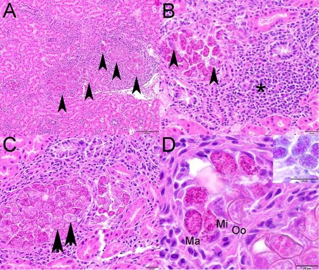
Figure 1. Photomicrographs from a Canada Goose (Branta canadensis) from Ohio, USA. Hematoxylin and eosin stain. (A) Renal tubules are distended with various stages of intraepithelial coccidia (arrowheads) and are surrounded by inflammation. (B) Macrogamonts and fewer microgamonts (arrowheads) expand the cytoplasm of renal tubular epithelial cells and the affected tubule is surrounded by large numbers of lymphocytes and plasma cells and fewer macrophages and granulocytes (asterisk). (C) Large numbers of coccidial oocysts (arrowheads) fill the cytoplasm of renal tubular epithelial cells. (D) Macrogamonts (Ma) with peripheral PAS positive granules (inset), microgamonts (Mi), and unsporulated oocysts with a thick wall (Oo) are present in the cytoplasm of renal tubular epithelial cells.
Disease: Renal coccidiasis refers to infection while coccidiosis refers to infections resulting in clinical disease. In this case, the finding of coccidia in the kidney was incidental.
Etiology: Most infections in birds are caused by Eimeria spp. Geese are commonly infected with E. truncata.
Host range: Infections have been reported in a wide variety of avian species, including Anseriformes, Apterygiformes, Charadriiformes, Gaviiformes, Procellariiformes, Sphenisciformes, Strigiformes, and Suliformes. Young birds or birds stressed by other conditions are more susceptible.
Distribution: Worldwide.
Seasonality: Occurs more commonly when birds are aggregated for breeding or in wintering grounds.
Life cycle: The life cycle is direct. An infected bird sheds oocysts in feces. Within one to five days, the sporont within the oocyst divides to form four sporocysts. Each sporocysts contains two infective sporozoites. A bird host ingests the infective oocyst and the sporozoites invade the renal epithelial cells where they undergo sexual reproduction to produce unsporulated oocysts which are shed in the feces.
Transmission: Infection occurs through the ingestion of material contaminated with infectious oocysts.
Clinical signs: Cases are often asymptomatic. Young birds may be weak or emaciated.
Gross findings:
- Emaciation and pectoral muscle atrophy
- Severe infections – enlarged kidneys with multifocal to coalescing tan foci
Microscopic findings: The architecture of renal tubules is distorted with dilated tubules with swollen cells that contain intracellular coccidian stages. There may be necrosis of renal tubular epithelial cells. Affected tubules are often surrounded by inflammatory cells.
Diagnosis: Gross and microscopic findings, direct smears of kidney; direct examination of oocysts or flotation.
Immunity: Domestic geese develop immunity to reinfection with E. truncata.
Wildlife population impact: While outbreaks may involve hundreds of birds, there is likely no impact on population health.
Public health concerns: None.
Management: Control is not feasible.
References:
United States Geological Survey. 1999. Field manual of wildlife diseases: general field procedures and diseases of birds, https://pubs.er.usgs.gov/publication/itr19990001. Accessed April 2025.
Yabsley MJ. 2008. Eimeria. In: Parasitic Diseases of Wild Birds, Atkinson CT, Thomas NJ, Hunter DB, editors. Wiley-Blackwell, Ames, Iowa, pp 162–180.
Related

WHISPers

Pathology Case of the Month

Diagnostic Services
Related
WHISPers
Pathology Case of the Month