
Acute renal failure in a Laughing Gull
History: A 280 gram adult Laughing Gull (Leucophaeus atricilla) was found dead in Vermont, USA. This gull was part of a mortality event involving fifteen gulls of several different species. One sick gull was observed standing but listless and did not flee when approached.
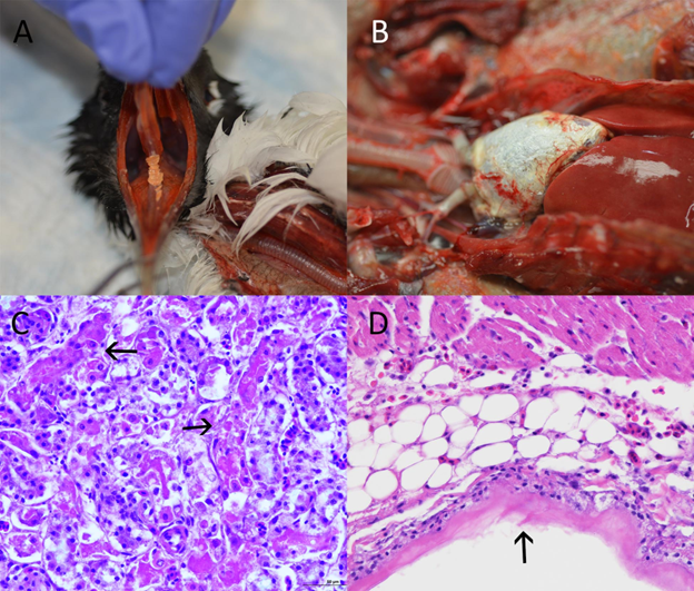
Gross Findings: On external examination, the keel is prominent. A large (approximately 1.5 x 0.3 cm) raised caseous plaque is present on the oral mucosa beneath the tongue (Fig 1A). Scattered smaller plaques are present on the pharyngeal mucosa. On internal examination, there is scant subcutaneous and visceral fat. The pericardium is diffusely thickened by dry, gritty, white material (Fig 1B) and is adhered to the epicardial surface. Similar material is present in multiple foci on the serosal surfaces of the spleen, liver, and trachea. The kidneys are pale and swollen.
Histopathological Findings: In the kidney, there is multifocal, moderate to marked, acute necrosis of the proximal tubular epithelial cells. Necrotic cells have pyknotic, karyorrhectic, or missing nuclei, and the lumens of affected tubules contain sloughed, brightly eosinophilic, necrotic cellular debris (Fig 1C). Urate tophi are present within multiple distal tubules. No crystals are seen under polarized light. In the oral cavity, there are multiple mucosal ulcerations covered by mats of necrotic debris. Hemorrhages are present in the subcutaneous tissue and skeletal muscle underlying the ulcerated areas, accompanied by multifocal to locally extensive, moderate heterophilic infiltrates. The pericardial surface is diffusely covered by amorphous, somewhat granular material consistent in appearance with uric acid deposits (Fig 1D). Multifocal, mild infiltrates of heterophils and scattered fibrin strands are present within the epicardial fat and the epicardium.
Morphologic Diagnoses:
- Kidney - Acute, marked, multifocal proximal epithelial tubular necrosis
- Pericardium - Acute, severe, diffuse uric acid deposition (gout)
- Oropharyngeal mucosa – Acute, moderate, multifocal ulceration
Disease: Acute renal failure
Etiology: The cause of the acute renal failure (ARF) seen in this gull was not determined. The principle causes of acute proximal tubular necrosis in birds and other animals are ischemia or toxicosis. Ischemia may cause ARF in wild birds which suffer from dehydration, shock, and other conditions leading to poor renal perfusion. In the case of a die-off of multiple wild birds, as was seen in this case, exposure to a nephrotoxic compound is usually suspected. A GC/MS toxicology screen using gastrointestinal content from this gull was negative; none of the toxic organic compounds that can be detected by this test were present. Other nephrotoxins that may cause ARF in birds include but are not limited to some mycotoxins, metals, plants, and animal venoms.
Distribution/Seasonality: As noted above, there are many different potential causes of ARF in wild birds, and therefore seasonality and spatial distribution of this condition are not specified.
Host Range: Due to the many potential causes of ARF, any species of wild bird may be affected. However, scavenging birds, rock doves (Columba livia) and various species of blackbirds may be overrepresented in cases of nephrotoxicosis. For example, scavengers may consume toxic substances in discarded carcasses, as was the case in mass die-offs of White-rumped vultures (Gyps bengalensis) in India and Pakistan due to consumption of diclofenac. Rock doves and blackbirds may be targeted as “nuisance birds” and intentionally poisoned by bait containing nephrotoxic avicides.
Transmission: Causes of ischemic ARF are many and modes of transmission are not generally described. Transmission of nephrotoxins to birds is most often due to ingestion of a poisonous substance.
Clinical Signs: Anorexia, lethargy, polydipsia, polyuria, and progressive weight loss are commonly seen.
Pathology: Ischemic events or exposure to nephrotoxins that target the proximal tubular epithelial cells result in destruction of the nephron. If the insult is severe enough, the kidney is no longer able to function sufficiently. Gout is the result of blocked excretion of uric acid by the kidney, and deposition on multifocal serous membrane surfaces of the viscera, often including the kidney, heart, liver, mesentery, air sac and peritoneum. High levels of uric acid in the bloodstream may also lead to ulcerative stomatitis.
Diagnosis: Antemortem diagnosis of ARF in wild birds can be challenging, as many of the clinical signs are non-specific. Gross necropsy findings of swollen pale kidneys and visceral gout are highly suggestive of ARF, which can be confirmed by histopathologic examination of the kidneys.
Public Health Concerns: Direct public health concerns of ARF in wild birds are determined by the cause. If nephrotoxins are present in the environment, accidental ingestion by humans is theoretically possible. In the case of the mass poisoning of vultures in India and Pakistan, ARF was an indirect cause of human health issues. The loss of approximately 95% of the vulture population due to diclofenac poisoning led to a significant increase in livestock carcasses on the landscape that the vultures would normally have consumed. Increased numbers of feral dogs were drawn to these livestock carcasses and the dense population of these dogs led to outbreaks of canine rabies. Cases of human rabies then followed due to an increase in bites from rabid dogs.
Wildlife Population Impacts: Impacts to wild bird populations due to ARF are likely highly variable. Exposure to nephrotoxins may affect only a few birds or may lead to significant mass die-offs as discussed above.
Management: In situations where avian mortality due to known nephrotoxin exposure occurs, removal of the toxin from the environment is an effective intervention.
References:
Fulton RM. 2020. Toxins and Poisons. In Diseases of Poultry (eds Swayne DE, Boulianne M, Logue CM, et. al.). https://doi.org/10.1002/9781119371199.ch32
Lierz M. 2003. Avian renal disease: pathogenesis, diagnosis, and therapy. Vet Clin North Am Exot Anim Pract 6(1):29-55. doi: 10.1016/s1094-9194(02)00029-4. PMID: 12616833.
Meteyer CU, Rideout BA, Gilbert M, Shivaprasad HL, Oaks JL. 2005. Pathology and proposed pathophysiology of diclofenac poisoning in free-living and experimentally exposed Oriental White-backed vultures (Gyps bengalensis). J Wild Dis 41(4):707-716. doi:10.7589/0090-3558-41.4.707.
Oaks J, Gilbert M, Virani M. et al. 2004. Diclofenac residues as the cause of vulture population decline in Pakistan. Nature 427:630–633. https://doi.org/10.1038/nature02317
Schmidt RE. 2006. Types of renal disease in avian species. Vet Clin North Am Exot Anim Pract 9(1):97-106. doi: 10.1016/j.cvex.2005.10.003. PMID: 16407081; PMCID: PMC7110740.
Schoemaker, NJ and Redig PT. 1997. Visceral gout in two red‐tailed hawks (Buteo jamaicensis). Veterinary Quarterly, 19(sup 1):61–62. https://doi.org/10.1080/01652176.1997.9694818
Sobhakumari A, Poppenga RH, Tawde S. 2018. Avian Toxicology. In Veterinary Toxicology (Third Edition). Gupta RC, (ed). Academic Press. ISBN 9780128114100, https://doi.org/10.1016/B978-0-12-811410-0.00053-2.
Related

WHISPers

Pathology Case of the Month

Diagnostic Services
Related
WHISPers
Pathology Case of the Month