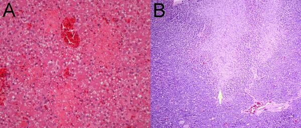
Photomicrographs from a sea otter (Enhydra lutris) found dead in Washington State, USA.
Valerie Shearn-Bochsler
Valerie Shearn-Bochsler is a Wildlife Pathologist at the National Wildlife Health Center.
Science and Products
Necropsy & Pathology
The Necropsy and Pathology services are performed by board-certified veterinary pathologists and necropsy technical staff whose principal role is to determine the cause of death for animals submitted to the USGS National Wildlife Health Center.
Epidemiologic and diagnostic data from two red foxes (Vulpes vulpes) and one fisher (Pekania pennanti) with highly pathogenic avian influenza H5N1 submitted to the USGS National Wildlife Health Center in 2022
Dataset: Acute oral toxicity and tissue residues of saxitoxin in the mallard (Anas platyrhynchos)
Data from pathology of Lagovirus europaeus GI.2/RHDV2/b (rabbit hemorrhagic disease virus 2) in native North American lagomorphs
Occurrence of Chlamydia sp in apparently healthy birds associated with a mortality event caused by chlamydiosis: Data
West Nile virus susceptibility of American singer canaries: Data
Dataset: Mortality trends in the Washington population of northern sea otters (Enhydra lutris kenyoni) during 20022015
Susceptibility and antibody response of the laboratory model zebra finch
Photomicrographs from a sea otter (Enhydra lutris) found dead in Washington State, USA.
Photographs and photomicrographs from a Common Raccoon (Procyon lotor) found dead in Wisconsin, USA. (A) There is green-gray mucoid discharge around the eyes and the nasal planum is crusty (arrows).
Photomicrographs from a Mexican Wolf (Canis lupus baileyi) euthanized due to neurologic illness in New York, USA. (A) Most of the neoplasm consists of loosely arranged, highly vacuolated stellate cells within a variably fine fibrovascular stroma. (B) An area of necrosis (asterisk) surrounded by pseudopalisading neoplastic cells (arrows).
Photographs from a Mexican Wolf (Canis lupus baileyi) euthanized due to neurologic illness in New York
linkPhotographs from a Mexican Wolf (Canis lupus baileyi) euthanized due to neurologic illness in New York, USA. (A) An area of light gray discoloration is present on the surface of the left cranio-dorsal cerebrum (arrow).
Image from a Timber Wolf found dead in Northern Wisconsin, USA showing round yeasts in necrotic debris within an area of osteomyelitis in the atlas
linkImage from a Timber Wolf (Canis lupus) found dead in Northern Wisconsin, USA. Multiple 8-20 micron, extracellular, round yeasts, some exhibiting broad-based budding, are present in necrotic debris within an area of osteomyelitis in the atlas (arrow) (markedly autolyzed tissue). Grocott’s Methenamine Silver (GMS) stain.
Atlas bone from Timber Wolf found dead in Wisconsin, USA showing multiple areas of lysis of bone
linkImage from a Timber Wolf (Canis lupus) found dead in Northern Wisconsin, USA. There are multiple areas of lysis of bone in the atlas (arrows).
Atlas bone from Timber Wolf found dead in Wisconsin, USA showing multiple areas of lysis of bone
linkImage from a Timber Wolf (Canis lupus) found dead in Northern Wisconsin, USA. There are multiple areas of lysis of bone in the atlas (arrows).
Image from a Timber Wolf (Canis lupus) found dead in Northern Wisconsin, USA. Ulceration of the mucosa of the distal esophagus (arrows). A plastic bag was extracted from where it was tightly lodged in the cardia of the stomach.
Image from a Timber Wolf (Canis lupus) found dead in Northern Wisconsin, USA. A focus of bony proliferation is seen on a radiograph of the left proximal radius (arrow).
Photographs and photomicrographs from a Great-horned Owl (Bubo virginianus) found dead in Arizona, USA
linkPhotographs and photomicrographs from a Great-horned Owl (Bubo virginianus) found dead in Arizona, USA. (A) Caseous material is present on the surface of the pharynx (arrows). (B) Multiple small tan foci are present in the liver, which is pale and moderately enlarged. (C) A focus of acute lytic necrosis in the liver (arrows). H&E stain.
Photographs and photomicrographs from a Common Raccoon (Procyon lotor) found dead in a prairie in Wisconsin, USA. (A) There is abrupt segmental reddening of the serosa of the distal jejunum and proximal ileum (arrows). (B) The mucosa of an affected area of the distal jejunum is reddened and bloody (C).
Liver from a Green Frog (Lithobates clamitans) tadpole. (A) Over 90% of the organ is infiltrated, effaced and replaced by protozoal spores (Perkinsea) admixed with necrotic debris. (B) Perkinsea spores are 4 to 6-µm in diameter spherical structures with thick, deep basophilic walls, and granular pale basophilic cytoplasm. H&E stain.
Gosner stage 41 Green Frog (Lithobates clamitans) tadpole. (A) There are no apparent external abnormalities. (B)The liver and spleen are markedly pale and enlarged.
Photographs and a radiograph from a Northern sea otter (Enhydra lutris kenyoni) from Oregon, U.S.A. (A) The deceased sea otter on a beach in Oregon, U.S.A.
Photomicrograph from a raccoon (Procyon lotor) found sick and euthanized in Texas, US. (A) A cluster of schistosome ova are present in the pancreas. Ova are thin-shelled and are not operculated. Each viable egg contains a miracidium (arrows). The ova are surrounded by infiltrates of eosinophils, macrophages, and lymphocytes. H&E stain.
Photographs from a red wolf (Canis rufus) found dead in North Carolina, USA. (A) Adult heartworms (Dirolfilaria immitis) in the right ventricle and atria of the heart and extending into the pulmonary artery. (B) Hard, haired nodule on the medial surface of the distal radius of the right leg.
Photomicrographs from a Common Eider (Somateria mollissima) from Massachusetts, USA. (A) Multiple pale foci of acute hepatic necrosis. (B) Multiple foci of acute pancreatic necrosis (arrow).
Photographs from a Common Eider (Somateria mollissima) from Massachusetts, USA. (A) There are scattered, small, pale foci in the liver. (B) Petechial and ecchymotic hemorrhages are present in the pancreas.
West Nile virus (avian) case definition for wildlife
Highly pathogenic avian influenza A(H5N1) virus clade 2.3.4.4b infections in wild terrestrial mammals, United States, 2022
Acute oral toxicity and tissue residues of saxitoxin in the mallard (Anas platyrhynchos)
Pathology of Lagovirus europaeus GI.2/RHDV2/b (rabbit hemorrhagic disease virus 2) in native North American lagomorphs
Quarterly wildlife mortality report January 2021
Investigation of the 2018 thick-billed murre (Uria lomvia) die-off on St. Lawrence Island rules out food shortage as the cause
Lethal infection of wild raptors with highly pathogenic avian influenza H5N8 and H5N2 viruses in the USA, 2014–15
High costs of infection: Alphavirus infection reduces digestive function and bone and feather growth in nestling house sparrows (Passer domesticus)
Chlamydia psittaci in feral Rosy-faced Lovebirds (Agapornis roseicollis) and other backyard birds in Maricopa County, Arizona
West Nile virus infection in American singer canaries: An experimental model in a highly susceptible avian species
Grass is not always greener: Rodenticide exposure of a threatened species near marijuana growing operations
Mortality trends in northern sea otters (Enhydra lutris kenyoni) collected from the coasts of Washington and Oregon (2002–15)
Science and Products
Necropsy & Pathology
The Necropsy and Pathology services are performed by board-certified veterinary pathologists and necropsy technical staff whose principal role is to determine the cause of death for animals submitted to the USGS National Wildlife Health Center.
Epidemiologic and diagnostic data from two red foxes (Vulpes vulpes) and one fisher (Pekania pennanti) with highly pathogenic avian influenza H5N1 submitted to the USGS National Wildlife Health Center in 2022
Dataset: Acute oral toxicity and tissue residues of saxitoxin in the mallard (Anas platyrhynchos)
Data from pathology of Lagovirus europaeus GI.2/RHDV2/b (rabbit hemorrhagic disease virus 2) in native North American lagomorphs
Occurrence of Chlamydia sp in apparently healthy birds associated with a mortality event caused by chlamydiosis: Data
West Nile virus susceptibility of American singer canaries: Data
Dataset: Mortality trends in the Washington population of northern sea otters (Enhydra lutris kenyoni) during 20022015
Susceptibility and antibody response of the laboratory model zebra finch
Photomicrographs from a sea otter (Enhydra lutris) found dead in Washington State, USA.
Photographs and photomicrographs from a Common Raccoon (Procyon lotor) found dead in Wisconsin, USA. (A) There is green-gray mucoid discharge around the eyes and the nasal planum is crusty (arrows).
Photomicrographs from a Mexican Wolf (Canis lupus baileyi) euthanized due to neurologic illness in New York, USA. (A) Most of the neoplasm consists of loosely arranged, highly vacuolated stellate cells within a variably fine fibrovascular stroma. (B) An area of necrosis (asterisk) surrounded by pseudopalisading neoplastic cells (arrows).
Photographs from a Mexican Wolf (Canis lupus baileyi) euthanized due to neurologic illness in New York
linkPhotographs from a Mexican Wolf (Canis lupus baileyi) euthanized due to neurologic illness in New York, USA. (A) An area of light gray discoloration is present on the surface of the left cranio-dorsal cerebrum (arrow).
Photographs from a Mexican Wolf (Canis lupus baileyi) euthanized due to neurologic illness in New York
linkPhotographs from a Mexican Wolf (Canis lupus baileyi) euthanized due to neurologic illness in New York, USA. (A) An area of light gray discoloration is present on the surface of the left cranio-dorsal cerebrum (arrow).
Image from a Timber Wolf found dead in Northern Wisconsin, USA showing round yeasts in necrotic debris within an area of osteomyelitis in the atlas
linkImage from a Timber Wolf (Canis lupus) found dead in Northern Wisconsin, USA. Multiple 8-20 micron, extracellular, round yeasts, some exhibiting broad-based budding, are present in necrotic debris within an area of osteomyelitis in the atlas (arrow) (markedly autolyzed tissue). Grocott’s Methenamine Silver (GMS) stain.
Image from a Timber Wolf found dead in Northern Wisconsin, USA showing round yeasts in necrotic debris within an area of osteomyelitis in the atlas
linkImage from a Timber Wolf (Canis lupus) found dead in Northern Wisconsin, USA. Multiple 8-20 micron, extracellular, round yeasts, some exhibiting broad-based budding, are present in necrotic debris within an area of osteomyelitis in the atlas (arrow) (markedly autolyzed tissue). Grocott’s Methenamine Silver (GMS) stain.
Atlas bone from Timber Wolf found dead in Wisconsin, USA showing multiple areas of lysis of bone
linkImage from a Timber Wolf (Canis lupus) found dead in Northern Wisconsin, USA. There are multiple areas of lysis of bone in the atlas (arrows).
Atlas bone from Timber Wolf found dead in Wisconsin, USA showing multiple areas of lysis of bone
linkImage from a Timber Wolf (Canis lupus) found dead in Northern Wisconsin, USA. There are multiple areas of lysis of bone in the atlas (arrows).
Image from a Timber Wolf (Canis lupus) found dead in Northern Wisconsin, USA. Ulceration of the mucosa of the distal esophagus (arrows). A plastic bag was extracted from where it was tightly lodged in the cardia of the stomach.
Image from a Timber Wolf (Canis lupus) found dead in Northern Wisconsin, USA. A focus of bony proliferation is seen on a radiograph of the left proximal radius (arrow).
Photographs and photomicrographs from a Great-horned Owl (Bubo virginianus) found dead in Arizona, USA
linkPhotographs and photomicrographs from a Great-horned Owl (Bubo virginianus) found dead in Arizona, USA. (A) Caseous material is present on the surface of the pharynx (arrows). (B) Multiple small tan foci are present in the liver, which is pale and moderately enlarged. (C) A focus of acute lytic necrosis in the liver (arrows). H&E stain.
Photographs and photomicrographs from a Great-horned Owl (Bubo virginianus) found dead in Arizona, USA
linkPhotographs and photomicrographs from a Great-horned Owl (Bubo virginianus) found dead in Arizona, USA. (A) Caseous material is present on the surface of the pharynx (arrows). (B) Multiple small tan foci are present in the liver, which is pale and moderately enlarged. (C) A focus of acute lytic necrosis in the liver (arrows). H&E stain.
Photographs and photomicrographs from a Common Raccoon (Procyon lotor) found dead in a prairie in Wisconsin, USA. (A) There is abrupt segmental reddening of the serosa of the distal jejunum and proximal ileum (arrows). (B) The mucosa of an affected area of the distal jejunum is reddened and bloody (C).
Liver from a Green Frog (Lithobates clamitans) tadpole. (A) Over 90% of the organ is infiltrated, effaced and replaced by protozoal spores (Perkinsea) admixed with necrotic debris. (B) Perkinsea spores are 4 to 6-µm in diameter spherical structures with thick, deep basophilic walls, and granular pale basophilic cytoplasm. H&E stain.
Gosner stage 41 Green Frog (Lithobates clamitans) tadpole. (A) There are no apparent external abnormalities. (B)The liver and spleen are markedly pale and enlarged.
Photographs and a radiograph from a Northern sea otter (Enhydra lutris kenyoni) from Oregon, U.S.A. (A) The deceased sea otter on a beach in Oregon, U.S.A.
Photomicrograph from a raccoon (Procyon lotor) found sick and euthanized in Texas, US. (A) A cluster of schistosome ova are present in the pancreas. Ova are thin-shelled and are not operculated. Each viable egg contains a miracidium (arrows). The ova are surrounded by infiltrates of eosinophils, macrophages, and lymphocytes. H&E stain.
Photographs from a red wolf (Canis rufus) found dead in North Carolina, USA. (A) Adult heartworms (Dirolfilaria immitis) in the right ventricle and atria of the heart and extending into the pulmonary artery. (B) Hard, haired nodule on the medial surface of the distal radius of the right leg.
Photomicrographs from a Common Eider (Somateria mollissima) from Massachusetts, USA. (A) Multiple pale foci of acute hepatic necrosis. (B) Multiple foci of acute pancreatic necrosis (arrow).
Photographs from a Common Eider (Somateria mollissima) from Massachusetts, USA. (A) There are scattered, small, pale foci in the liver. (B) Petechial and ecchymotic hemorrhages are present in the pancreas.