
Poxviral-associated epithelial neoplasia in a Black Tern.
History: An adult, male Black Tern (Chlidonias niger) was found unable to fly due to a large mass on the left wing. It was admitted to a rehabilitation center in Wisconsin, USA and died in care.
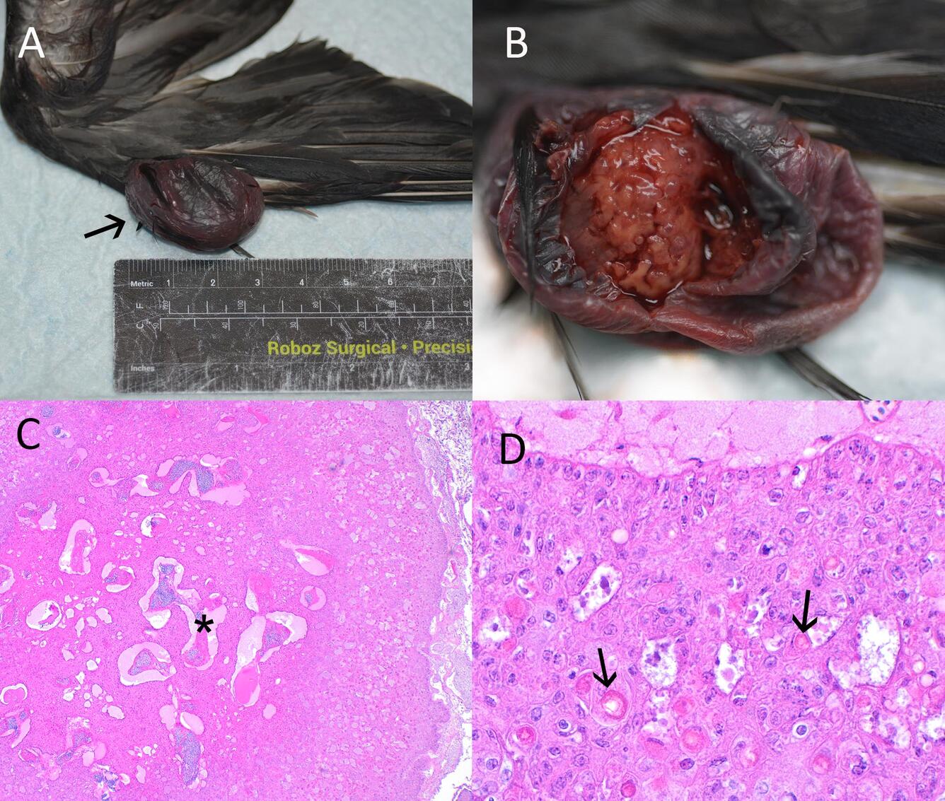
Gross Findings: A soft, fluctuant, roughly spherical, 3.5 x 2 x 1.5 cm cystic mass was present over the left metacarpus containing blood and irregular, firm, orange material (Fig. 1A, B). Overlying skin was mostly smooth with a few feathers protruding from the mass, and blood was present on the left wing. No subcutaneous, visceral or epicardial fat was present, and the pectoral muscles were atrophic and concave. Lungs were diffusely light red, wet, and floated in formalin. The ventriculus contained small fish bones. Intestines and pancreas were markedly autolytic. The liver was tan to light red, and the kidneys were pale tan with a pronounced reticular pattern. Gonads were not observed. Thymus and bursa were absent. The thyroid was light red, and the brain was pale and very soft.
Histopathological Findings: The mass on the wing was an unencapsulated, expansile mass composed of cords, nests and sheets of large, pleomorphic polygonal epithelial cells supported by a loose fibrovascular stroma around irregular vasculature and blood-filled spaces. Thick cords of neoplastic cells lined multilocular, variably sized spaces containing blood and clumps of necrotic, keratinized debris (Fig. 1C). Neoplastic cells had distinct cell margins with cellular bridges, abundant, finely stippled to vacuolated eosinophilic cytoplasm, and frequently contained large, brightly eosinophilic cytoplasmic inclusions that often peripheralized the nucleus (Bollinger bodies) (Fig. 1D). Keratinization and asynchronous maturation were present. Nuclei were large and oval to indented or crescent-shaped, with open chromatin and prominent, occasionally multiple nucleoli. Cells were rarely binucleate, and there was fewer than one mitotic figure per 400x field. Overlying epidermis was thin, with few adnexa.
Sources/Usage: Public Domain, USGS.
Gross and Morphologic Diagnosis: Skin and subcutis. Poxviral carcinoma of epidermal or feather follicle origin (squamous cell carcinoma or keratoacanthoma).
Disease: Avian pox with neoplastic transformation of epithelial cells.
Etiology: Avipoxvirus with neoplastic transformation.
Distribution: Avian poxviruses have worldwide distribution.
Seasonality: Pox can be seen year-round but acute poxviral infection may be more prevalent during warm weather and breeding seasons when insects are active and bird to bird contact is increased. Neoplastic sequalae are uncommon and not seasonal.
Host range: Avian pox is a worldwide group of viruses known to infect over 200 species of birds (Gyuranecz et al. 2013).
Transmission: Avipoxviruses are spread most effectively mechanically by mosquitoes and biting insects but can also be transmitted by aerosols, contact with contaminated surfaces, and direct contact between birds. Bird feeders can act as a source of transmission due to both increased congregation of birds and contamination of the feeder itself (Hansen 1999).
Clinical signs: The most commonly observed form of avian pox consists of raised, nodular growths seen on unfeathered skin and around the eyes and beak. Emaciation and open mouth breathing may be present when lesions impair feeding or obstruct airways, but birds able to feed may recover. Less common and seen in young birds, the diphtheritic form results in weakness, inability to eat, and difficulty breathing with necrotic plaques in the mouth, esophagus, and upper respiratory membranes. This form has a high mortality (Hansen 1999). Poxvirus-induced dermal squamous cell carcinoma and avian keratoacanthoma are seen sporadically (Pesaro et al. 2009). Latent poxviral infections are possible (Fallavena et al. 2002).
Pathology: Pox viruses express growth-factor homologues that induce mitosis within epithelial cells, creating the characteristic raised (nonneoplastic) lesions, and are associated with immunosuppression (Fallavena et al. 2002). They are associated with squamous cell carcinoma in several avian species, including wild and domestic birds (Fallavena et al. 2002, Pesaro et al. 2009, Braga et al. 2020, Mohamed et al. 2024). Several poxviruses of wild and domestic birds also carry integrated proviral sequences of reticuloendothelial virus, a retrovirus associated with neoplasia of lymphoid and other tissues (Giotis and Skinner 2019, Mosad et al. 2020). Both oncogenes within the virus and immunosuppression are likely to contribute to the formation of squamous cell carcinoma in affected birds.
Diagnosis: Histology showing characteristic large eosinophilic cytoplasmic inclusion bodies within epithelial cells and Immunohistochemistry or PCR of either acute (nonneoplastic) lesions or neoplastic masses. Typical gross lesions with PCR may be sufficient in acute infection.
Public health concerns: None.
Wildlife population impacts: Pox viral neoplasia is an uncommon sequela with minimal population impact, but acute pox viral epidemics can be responsible for marked morbidity (Senar and Conroy 2004) and detrimental to vulnerable populations, especially isolated or naive species (Van Riper et al. 2002).
Management: Mosquito control by eliminating standing water for breeding and discouraging bird congregation by removing feeders and bird baths for at least two weeks can help to reduce spread during an epidemic. Disinfection of bird feeders or baths with a 10% bleach solution before re-use deactivates residual pox virus (Cornell Wildife Health Lab 2006).
References:
Braga, J. F. V., R. M. Couto, M. C. Rodrigues, R. Ecco. 2020. Avipoxvirus detected in tumor-like lesions in a white-faced whistling duck (Dendrocygna viduata). Pesquisa Veterinária Brasileira 40: 818-823.
Cornell Wildlife Health Lab. 2006. Avian Pox. NYS Wildlife Health Program. https://cwhl.vet.cornell.edu/resource/avian-pox. (Accessed march 5, 2026)
Fallavena, L. C. B., C. W. Canal, C. T. P. Salle, H. L. S. Moraes, S. L. S. Rocha, R. A. Pereira, A. B. D. Silva. 2002. Presence of avipoxvirus DNA in avian dermal squamous cell carcinoma. Avian Pathology 31: 241-246.
Giotis, E. S., M. A. Skinner. 2019. Spotlight on avian pathology: fowlpox virus. Avian Pathology 48: 87-90.
Gyuranecz, M., J. T. Foster, Á. Dán, H. S. Ip, K. F. Egstad, P. G. Parker, J. M. Higashiguchi, M. A. Skinner, U. Höfle, Z. Kreizinger, G. M. Dorrestein, S. Solt, E. Sós, Y. J. Kim, M. Uhart, A. Pereda, G. González-Hein, H. Hidalgo, J.-M. Blanco, K. Erdélyi. 2013. Worldwide Phylogenetic Relationship of Avian Poxviruses. Journal of Virology 87: 4938-4951.
Hansen, W. 1999. Avian pox in Field manual of wildlife diseases: General field procedures and diseases of birds. M. Friend, J. C. Franson, (eds.) Information and Technology Report. U. S. Geological Survey, Reston, VA. 163-169 pp.
Mohamed, R. I., H. A. Elsamadony, R. A. Alghamdi, A. Eldin, A. El-Shemy, S. Abdel-Moez Amer, S. M. A. Bahshwan, M. T. El-Saadony, H. S. El-Sayed, K. A. El-Tarabily, A. S. A. Saad. 2024. Molecular and pathological screening of the current circulation of fowlpox and pigeon pox virus in backyard birds. Poultry Science 103: 104249.
Mosad, S. M., M. El-Tholoth, A. A. El-Kenawy, L. J. M. Abdel-Hafez, F. A. El-Gohary, H. El-Sharkawy, M. M. Elsayed, A. A. Saleh, E. K. Elmahallawy. 2020. Molecular Detection of Reticuloendotheliosis Virus 5′ Long Terminal Repeat Integration in the Genome of Avipoxvirus Field Strains from Different Avian Species in Egypt. Biology 9: 257.
Pesaro, S., B. Biancani, G. Fabbrizi, G. Rossi. 2009. Squamous cell carcinoma with presence of poxvirus-like inclusions in the foot of a pink-backed pelican (Pelecanus rufescens). Avian Pathology 38: 229-231.
Senar, J. C., ANDM. J. Conroy. 2004. Multi-state analysis of the impacts of avian pox on a population of Serins (Serinus serinus): The importance of estimating recapture rates. Animal Biodiversity and Conservation 27: 133-146.
van Riper, C.,III, S. G. van Riper, W. R. Hansen. 2002. Epizootiology and Effect of Avian Pox on Hawaiian Forest Birds. The Auk 119: 929-942.
Related

WHISPers

Pathology Case of the Month

Diagnostic Services
Related
WHISPers
Pathology Case of the Month