
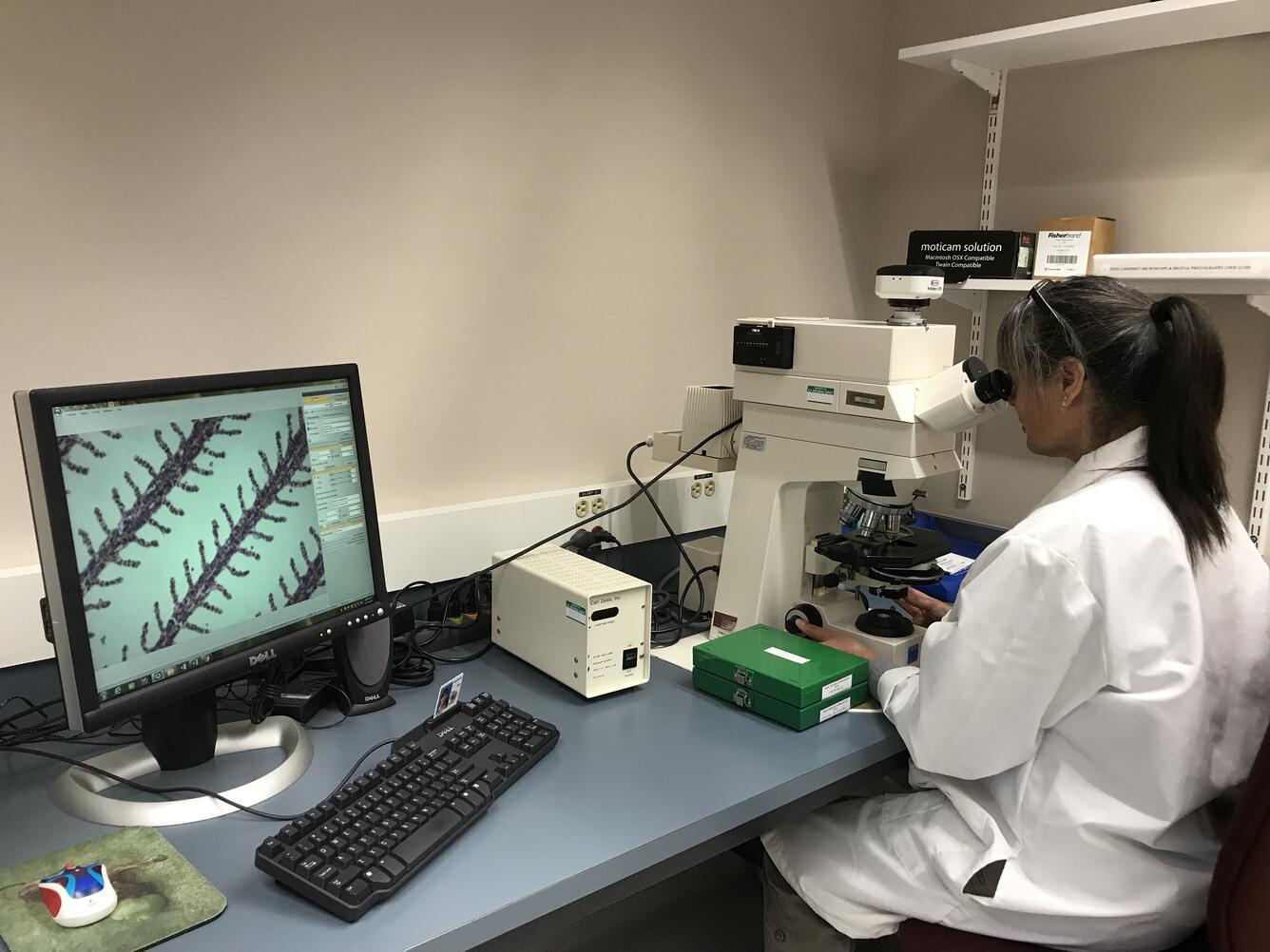
Histology Laboratory WFRC - Scientist at Microscope
Detailed Description
Tissue sections are mounted on glass slides, stained and examined with a microscope that magnifies cellular details up to 2,000 times with brightfield or fluorescence imaging. Microscopes are used in our research to understand the pathological changes caused by infectious agents such as bacteria, fungi, parasites and viruses. Additionally, microscopic abnormalities due to environmental conditions, contaminants and toxins can be detected and used to evaluate water quality.
Sources/Usage
Public Domain.