
Histopathology - Plate 21 - Figures 61-63 thumb
Detailed Description
Histopathology - Plate 21 - Figures 61-63
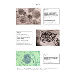
- Figure 61: N. salmonis. Kidney imprint of fingerling Chinook salmon infected with N. salmonis. (Courtesy of John Morrison).
- Figure 62: N. salmonis. Developing sporoblast (arrows) within the host cell nucleus of a fingerling Chinook salmon. (Courtesy of John Morrison).
- Figure 63: Loma sp. a microsporidian, in a luamellar cyst of a 2-year-old steelhead trout.