
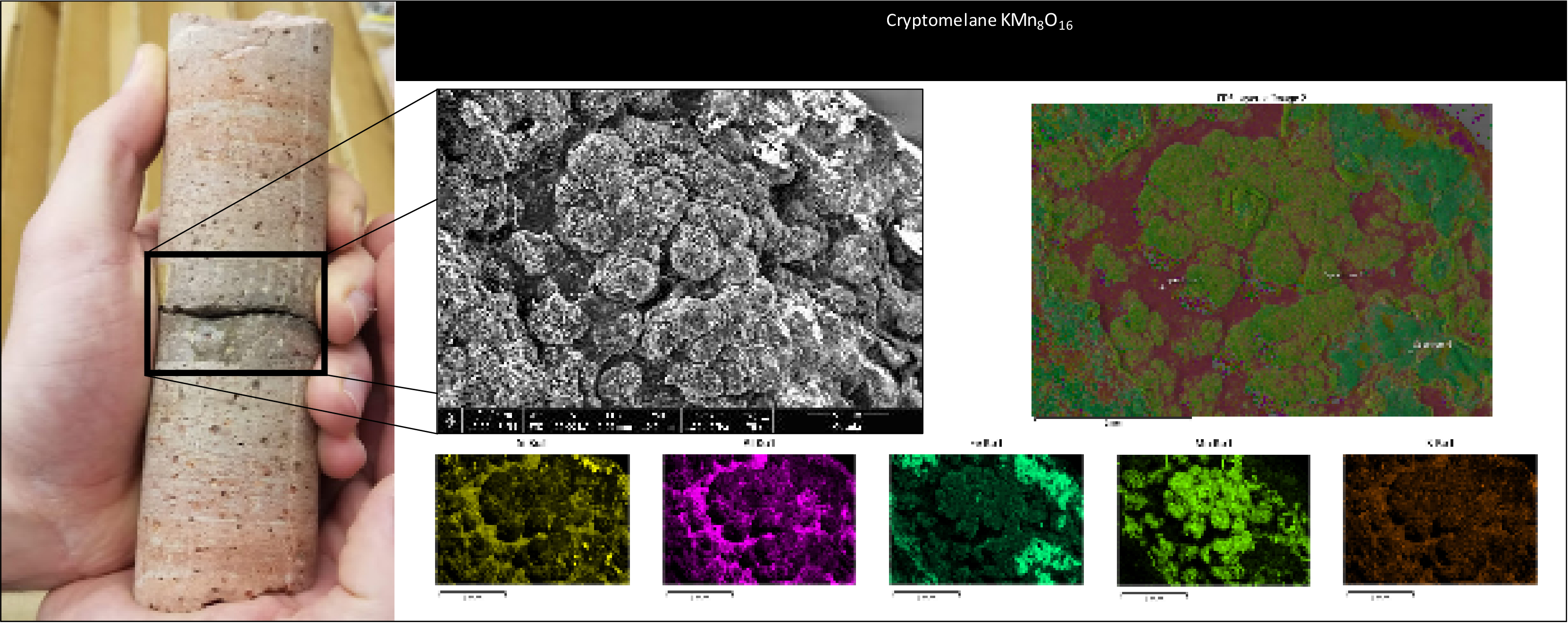
Scanning electron microscope image of Yellowstone drill core

Detailed Description
The right side of the figure is an image of a small piece of the Y-9 core from the USGS 1967-68 drilling expedition to Yellowstone National Park. The black area was analyzed using a scanning electron microscope (SEM) at the University of Wyoming to determine mineralogy and dispersion of elements. The black and white photo is an SEM image of the alteration mineral cryptomelane. The colored image is an SEM elemental distribution map. The smaller colored images are SEM element maps showing the isolated distributions of silicon, aluminum, iron, manganese, and potassium increasing in abundance from left to right.
Sources/Usage
Public Domain.