
Esophageal-proventricular Aspergillosis in a Canada Goose (Branta canadensis) from Wisconsin, USA
Canada Goose on the Colville River Delta
History: An adult male 2850-g Canada Goose (Branta canadensis) was observed lethargic, easily approachable, and with a tucked head in September in Wisconsin, USA. It was later found dead and submitted for necropsy.
Gross Findings: There were no significant external findings. On internal examination, no subcutaneous, visceral or epicardial fat was observed (emaciation). Multiple green plaques were present on the dorsal surface of the keel, the pericardial sac, and on the thickened cranial and caudal air sacs. Lungs were diffusely red and contained numerous, variably sized, tan nodules on cut section. At the junction of the esophagus and proventriculus, there were five raised tan nodules with a central depression. Nodules measured approximately 3 mm in diameter by 2 mm (Fig. 1A, B)
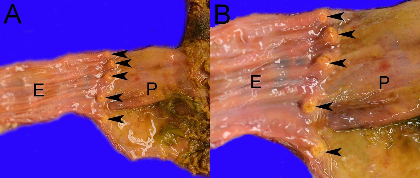
Figure 1. Photographs from a Canada Goose (Branta canadensis) found dead in Wisconsin, USA. (A) Five raised, tan nodules with a central depression (arrowheads) are present at the junction of the esophagus (E) and proventriculus (P). (B) Higher magnification showing the approximately 3 mm in diameter by 2 mm raised, tan nodules (arrowheads) at the junction of the esophagus (E) and proventriculus (P).
Histopathological Findings: At the junction of the esophagus and proventriculus, there are two granulomas (Fig. 2A). Granulomas have a core of eosinophilic debris admixed with numerous degenerate heterophils surrounded by epithelioid macrophages, further rimmed by lymphocytes and plasma cells, and connective tissue. Grocott's Methenamine Silver (GMS) stain positive, parallel walled fungal hyphae, with dichotomous, acute-angle branching are present in the core of the granuloma (Fig. 2B).
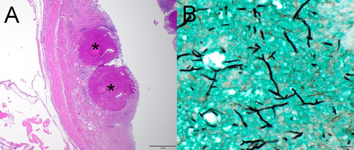
Figure 2. Photomicrographs from a Canada Goose (Branta canadensis) found dead in Wisconsin, USA. (A) Two granulomas (asterisks) are present at the junction of the esophagus and proventriculus. (B) Parallel walled fungal hyphae with dichotomous, acute-angle branching are present in the core of the granuloma (Fig. 2B). Grocott's Methenamine Silver stain.
Disease: Aspergillosis
Etiology: Aspergillus fumigatus: A ubiquitous saprophytic fungus found in soil, decomposing organic matter, agricultural waste grains, silage, litter, and moldy feed. Aspergilli grow well in moist environments at 23–26 °C. A. fumigatus is the most pathogenic species for wild birds.
Host range: Infections have been reported in a wide variety of avian species, especially in Anseriformes, Accipitriformes, Charadriiformes, Passeriformes, and Galliformes. Young birds or birds stressed by other conditions are more susceptible.
Distribution: Worldwide, including Antarctica.
Seasonality: In North America, outbreaks in waterfowl occur most often in the fall and early winter but can occur at any time. The fungi multiply in wet conditions and produce spores that disperse during dry conditions.
Pathogenesis: Spores are inhaled and germinate in the respiratory tract producing plaques of hyphae. Hyphae cause a strong inflammatory response with macrophages, multinucleated giant cells, and heterophils surrounding the hyphae. If hyphae produce conidia, they can germinate in other sites causing additional lesions. Hyphae are angioinvasive, and hematogenous dissemination can result in infection of any internal organ. While some species of Aspergillus can produce mycotoxins, the role these metabolites play in disease development is not clear.
Transmission: Infection occurs primarily through the inhalation of airborne spores, but can occur through puncture wounds, especially those that puncture air sacs.
Clinical signs: Acute infections may cause lethargy, dullness, ruffled feathers, dehydration, anorexia, isolation, gasping, bill opening and closing, dyspnea, and cyanosis. In chronic infections, clinical signs include emaciation, reduced activity, inability to fly, dyspnea or tachypnea, and vomiting and diarrhea. With invasion of the nervous system, clinical signs include ataxia, torticollis, or other neurological signs.
Gross findings:
- Acute infections – dark red edematous lungs with multiple small, tan nodules; firm, flat, yellow, cotton-like plaques in air sacs
Chronic infections – plaques in air sacs that may be dark-colored if conidia develop; lesions in any organ
Microscopic findings: Granulomas are most frequently observed in the respiratory tract, gastrointestinal tract, liver, spleen, kidney, eye, bone, or brain with characteristic parallel walled, septate hyphae with dichotomous, acute-angle branching. Hyphae stain with periodic acid-Schiff (PAS) reaction or GMS.
Diagnosis: History, clinical signs, gross and microscopic findings, culture, IHC, PCR.
Public health concerns: Aspergillosis is a zoonotic disease that causes pulmonary mycosis in humans, especially among those who are immunocompromised.
Wildlife population impacts: Aspergillosis can cause epizootics when birds that flock together are simultaneously exposed.
Management: When a mortality event occurs, plow under, bury or cover moldy feed, remove moldy feed from bird feeders, and keep nest boxes free of damp and moldy nesting material.
References:
Arné P, Risco-Castillo V, Jouvion G., Le Barzic C, & Guillot J. 2021. Aspergillosis in Wild Birds. J Fungi 7(3): 241. DOI:10.3390/jof7030241.
Converse KA. 2007. Aspergillosis. In: Infectious Diseases of Wild Birds, Thomas NJ, Hunter DB, Atkinson CT, editors. Blackwell Publishing, Ames, Iowa, pp 360–374.
Gonçalves VN, Amorim SS, da Costa MC, de Assis Santos D, Convey P, Rosa LH. 2024. Pathogenic potential of an environmental Aspergillus fumigatus strain recovered from soil of Pygoscelis papua (Gentoo penguins) colony in Antarctica. Braz J Microbiol 55(2):1521–1528. DOI:10.1007/s42770-024-01326-w.
Related

WHISPers

Pathology Case of the Month

Diagnostic Services
Related
WHISPers
Pathology Case of the Month