
Pathology Case of the Month - California Newt
History: In May 2021, a morbidity and mortality event involving California newts (Taricha torosa) was reported at a national park in California, USA.
Clinical signs of affected newts included cloudy eyes, a tail lesion, impaired righting reflex, and odd behavior including spinning or whirling. Newts with cloudy eyes, tail lesions, and poor body condition were previously reported in this area in 2020. In 2021, one emaciated newt was collected, euthanized, and submitted for diagnostic evaluation.
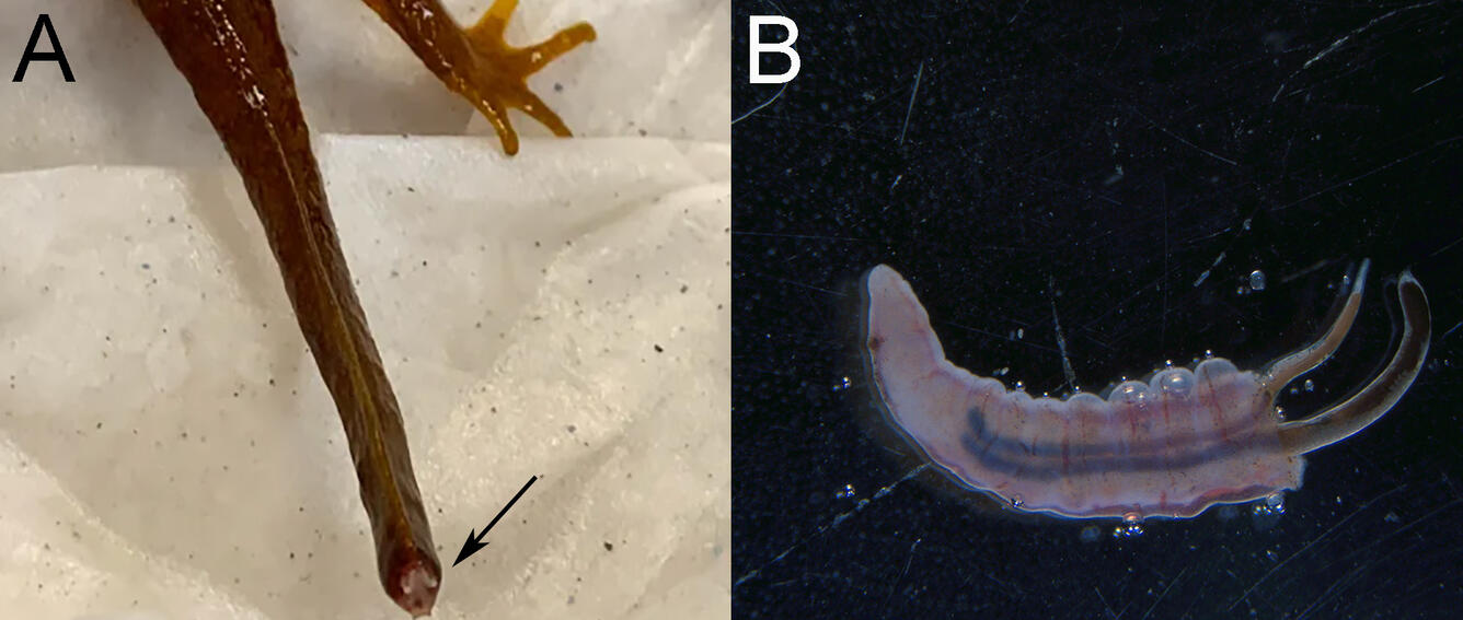
Gross Findings: The newt was in emaciated body condition, with prominent bony protuberances externally and no fat stores within the body. The eyes were clear and unremarkable. The tip of the tail was red with a 1 mm piece of vertebral bone protruding from the end (Fig. 1A). No other skin lesions were present. On internal examination, each lung contained 2-4 white to black lung worms ~9 mm in length within the lumen (Fig. 1B). There were no other significant internal findings.
Histopathological Findings: Microscopically, the tip of the tail had complete loss of skin and skeletal muscle with exposure of the vertebral bone (Fig. 2A). There was regrowth of hyperplastic epithelium along the edges of the wound, with moderate numbers of granular cells, mononuclear cells, and dilated lymphatics beneath the epithelium. There were moderate numbers of osteoclasts remodeling the exposed bone. There were a few superficial colonies of bacteria along the tip of the exposed bone (interpreted as not significant). Within the lumen of the lung were a few cross sections of Rhabditoid lung worms characterized by vacuolated lateral chords, an intestine containing dark pigment, and a uterus with developing larvae (Fig. 2B). There was minimal tissue reaction, with only segmental attenuation of the respiratory epithelium present. Within the stroma of the labyrinths of both middle ears, there was a moderate amount of edema and moderate numbers of mononuclear cells and rare granulocytes (Fig. 2C). Other microscopic findings included edema and inflammation in the intestines from an unknown cause, moderate renal tubular oxalosis likely secondary to ingestion of oxalate-rich plants in the environment, extramedullary hematopoiesis in the liver and spleen likely secondary to blood loss from the tail lesion, lymphoid depletion in the spleen, and moderate numbers of metacercariae (trematodes) primarily encysted in the wall of the intestines.

Diagnostic Test Results:
- Negative for Batrachochytrium dendrobatidis and B. salamandrivorans by PCR on skin swab
- Negative for Ranavirus by PCR on pooled liver and kidney
Morphologic Diagnosis/es:
- Emaciation
- Moderate bilateral mononuclear otitis media and edema
- Distal tail tip necrosis with bone exposure, epithelial regrowth, and bony remodeling
- Mild (colon) to moderate (small intestine) lymphoplasmacytic and histiocytic enterocolitis with segmental edema and few luminal nematodes and mucus
- Moderate multifocal renal tubular oxalosis
- Moderate multifocal encysted metacercariae (trematodes) within small and large intestines and liver
- Few Rhabditoid lung worms
Case Comment: This California newt had multiple comorbidities, including emaciation, distal tail tip necrosis, inflammation, and edema in both middle ears (likely cause of neurologic signs), pulmonary nematodiasis, renal oxalosis, and moderate parasitism in the gastrointestinal tract. The cause of neurologic signs was attributed to edema and inflammation in both middle ears. The underlying etiology/-ies for the tail tip necrosis and inflammation in the ears was/were not apparent and may be multifactorial, including water quality issues.
Disease: Pulmonary nematodiasis
Etiology: Rhabditida lung worm, suspect Rhabdias species based on location in the host and the host species.
Distribution: Worldwide
Host range: Rhabdias species are common parasites of both free-ranging and captive anuran (frogs and toads) and caudate (salamanders and newts) amphibians.
Life cycle: Rhabdias species have a direct life cycle. Reproductive stages occur both within the host and in the environment. Hermaphroditic adults in the lung produce eggs or larvae that are swallowed and pass through the gastrointestinal tract to be shed in feces. Larvae develop in the environment into male and female adults that mate to produce infective larvae that penetrate the skin of the amphibian host and migrate internally to the lung.
Clinical signs: Clinical signs range from none to anorexia, weight loss, wasting, skin disease, difficulty breathing, or sudden death.
Pathology: Rhabdias species are mottled white-black worms found within the lumen of the lung. There are no histologic lesions with low parasite loads. With heavy parasite burdens, there is pneumocyte hypertrophy and hyperplasia with mild interstitial infiltrates of lymphocytes, macrophages, or occasional eosinophils. Histologic features characteristic of Rhabdias spp. include scant platymyarian musculature, vacuolated lateral chords, a uterus with developing larvae, and an intestinal tract containing abundant dark pigmented material. Mixed inflammatory lesions can also be seen in the skin and soft tissues secondary to penetration of the larvae into the skin and migration to the lungs.
Diagnosis: Identification of morphologically compatible nematodes on gross examination or histopathology. Embryonated eggs or larvae can be recovered from fecal samples or tracheal wash specimens.
Public health concerns: None
Wildlife population impacts: There are over 40 different species of amphibian lung worm; many Rhabdias species are host-specific while others are generalists. Impacts to wildlife populations are likely low, though may be of concern for captive populations of amphibians where high loads of the parasite may accumulate in the environment.
Management: Because Rhabdias spp are transmitted through the environmental, treatment of wildlife populations is not practical. There are a variety of anthelmintics for use in captive species at various dosages for the treatment of individual animals. In general, two treatments are given two weeks apart, followed two weeks later by fecal examination. Treatments may be continued on this cycle until the parasites are cleared from the animal.
References:
- de la Navarre B. 2011. Common parasitic disease of reptiles & amphibians (Proceedings). https://www.dvm360.com/view/common-parasitic-diseases-reptiles-amphibians-proceedings. Accessed August 2021.
- Eisenberg T and Pantchev N. 2009. Infection with Rhabdias tokyoensis (Nematoda: Rhabdiasidae) in European captive-bred swordtail newts, Cynops ensicauda (Amphibia: Salamandridae). Salamandra 45(2):91-94.
- Hallinger MJ, Taubert A, and Hermosilla C. 2020. Endoparasites infecting exotic captive amphibian pet and zoo animals (Anura, Caudata) in Germany. Parasitology Research 119:3659-3763. https://doi.org/10.1007/s00436-020-06876-0
- Pessier AP. 2018. Amphibia. In: Pathology of Wildlife and Zoo Animals. Terio KA, McAloose D, St. Leger J, editors. Elsevier, San Diego, CA, pp. 941-942. https://doi.org/10.1016/B978-0-12-805306-5.00038-9
Related

WHISPers

Pathology Case of the Month

Necropsy & Pathology Necropsy & Pathology

Diagnostic Services
Related
WHISPers
Pathology Case of the Month
Necropsy & Pathology Necropsy & Pathology