
Pathology Case of the Month - Little Brown Bat
History: A Little Brown Bat (Myotis lucifugus) was captured in a harp trap in May 2023 as part of surveillance for Pseudogymnoascus destructans at a roost in Wyoming, U.S.A. Pseudogymnoascus destructans was documented at the site the previous year. No mortality was observed at the time of the survey. At the time of capture, nodules were observed on the patagia, and a skin biopsy was collected.
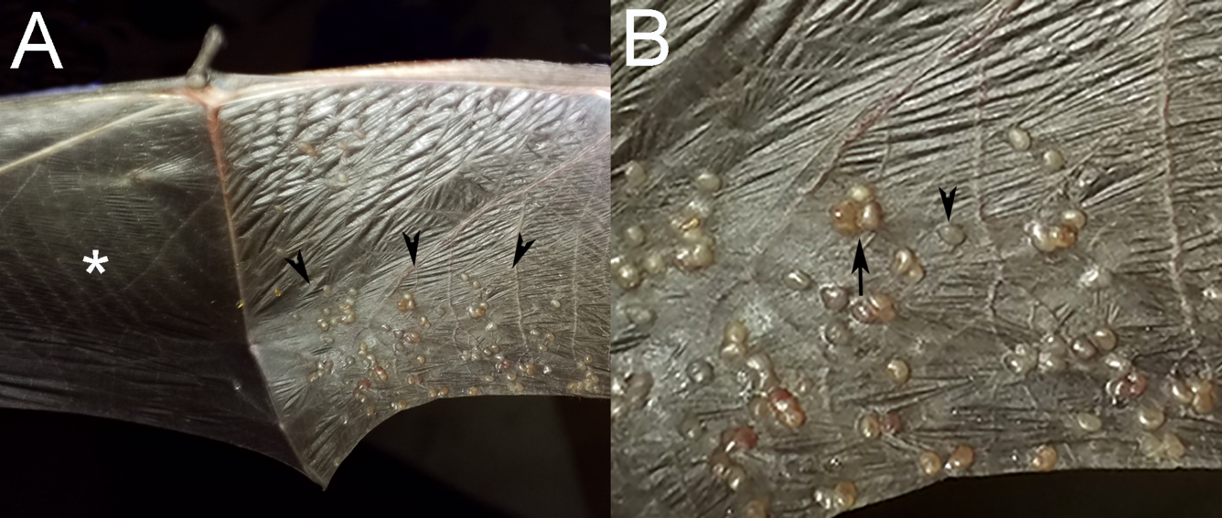
Gross Findings: On the caudoventral right plagiopatagium, there were numerous approximately 1–2 mm in diameter, irregularly round, slightly raised, vesicular, tan nodules that often occurred in clusters (Figs. 1A, B). Several mites were present on the patagium near the nodules.
Histopathology: Adult nematodes filled with larvae were present in the dermis (Fig. 2A). The overlying epidermis was hyperplastic. Nematodes were surrounded by low numbers of eosinophils, fewer lymphocytes and plasma cells, and fibroblasts (Fig. 2B). A few nematode larvae were observed on the surface of the epithelium (not pictured). No PAS positive fungal hyphae were observed.

Diagnostic test results: PCR-based tests for Pseudogymnoascus destructans (the causative agent of white-nose syndrome) and pox virus were negative.
Etiologic diagnosis: Cutaneous nematodiasis (suspect Muspiceoidea: Muspiceidae)
Etiology: There are five genera within the familiy Muspiceidae (Muspicea, Riouxgolvania, Lukonema, Pennisia, and Maseria) four of which have been observed in tissues of bats. Muspicea borreli occurs in subcutaneous tissues of mice in France, Germany and Australia. Four Riouxgolvania spp. have been described in the skin of bats.In 1958, Riouxgolvania rhinolophi was observed in the skin of the Mediterranean Horseshoe Bat (Rhinolophus euryale) from a cave in France. In 1973, R. nyctali was observed in a Common Noctule (Nyctalus noctula) in Holland and from an ear cyst in a Lesser Mouse-eared Bat (Myotis blythii; oxygnathus) from France. R. beveridgei was collected from the patagia of the Eastern Bent-wing Bat (Miniopterus schreibersi oceanensis) from Australia in 1978 and described in the Southern Bentwing Bat (Miniopterus schreibersii bassanii) collected in Australia in 2009. In 2007, R. kapapkamui was identified in dermal nodules in the Japanese Large-footed Bat (Myotis macrodactylus) and Ikonnikov's Bat (Myotis ikonnikovi) from Japan. Lukonema lukoschusi was observed in the uropatagium of bats in Surinam and French Guyana. Pennisia nagorseni was documented in the wings in bats from Kenya and Zimbabwe (formerly Rhodesia). Maseria vespertilionis was observed in the feet of bats from Oregon and Alaska.
Distribution: Muspiceidae have been documented in tissues of bats in France, Germany, Holland, Australia, Japan, Surinam, French Guyana, Kenya and Zimbabwe (formerly Rhodesia).
Transmission: Modes of transmission of the Muspiceidaare not well understood. Proposed mechanisms include mucocutaneous penetration, ingestion, cannibalism or transmission via hematophagous parasites. Close contact in bats in caves would allow spread through skin penetration.
Life cycle: Life cycles are unknown. Of the Muspiceidae, Pennisia nagorsenia is dioecious (male and female reproductive organs in separate individuals), while Muspicea borreli, Riouxgolvania spp., Lukonema lukoschusi, and Maseria vespertilionis are protandrous hermaphrodites, beginning life as males, and when needed are capable of transforming into females.
Gross findings: Dermal nodules that have been observed in the patagia of bats are white to tan, 1-2 mm in diameter, raised, vesicular, and may have a central crusted pore.
Histologic findings: Lesions are often granulomas centered on a nematode, but neutrophilic, eosinophilic and lymphocytic inflammation has also been observed. A crusted pore might be observed in the section. Co-infection with pox virus was noted in a single case.
Diagnosis: Histopathology, morphologic identification, PCR.
Public health concerns: Muspiceidae are not known to infect humans. Haycocknema perplexum (Robertdollfusiidae) occurs in myocytes of humans in Australia.
Wildlife population impacts: Cutaneous nematodiasis in bats is not considered to have a significant impact on wildlife population health.
Management: Management of this disease in wild bats is not practical.
References:
- Bain O, Chabaud A-G. 1968. Description de Riouxgolvania rhinolophi n. g., n. sp., nématode parasite de Rhinolophe, montrant les affinités entre Muspiceoidea et Mermithoidea. Ann Parasitol Hum Comp 43:45–50. https://doi.org/10.1051/parasite/1968431045
- Bain O, Chabaud A-G. 1979. Sur les Muspiceidae (Nematoda-Dorylaimina). Ann Parasitol Hum Comp 54:207–225. https://doi.org/10.1051/parasite/1979542207
- Chabaud AG, Bain O. 1974. Données nouvelles sur la biologie des Nématodes Muspicéides, fournies par l’étude d’un parasite de Chiloptères: Lukonema lukoschusi n. gen., n. sp. Ann Parasitol Hum Comp 48: 818–834.
- Hasegawa H, Satô M, Maeda K, Murayama Y. 2012. Description of Riouxgolvania kapapkamui Sp. N. (Nematoda: Muspiceoidea: Muspiceidae), a peculiar intradermal parasite of bats in Hokkaido, Japan. J Parasitol 98:995–1000. https://doi.org/10.1645/GE-2710.1
- McLelland DJ, Reardon T, Bourne S, Dickason C, Kessell A, Boardman W. 2013. Outbreak of skin nodules associated with Riouxgolvania beveridgei (Nematoda: Muspiceida) in the southern bentwing bat (Miniopterus schreibersii bassanii), South Australia. J Wildl Dis 49:1009–1013. https://doi.org/10.7589/2012-11-288
- Rausch RL, Rausch VR. 1983. Maseria vespertilionis n.g., n.sp. (Dorylaimina : Muspiceidae), a nematode from nearctic bats (Vespertilionidae). Ann Parasitol Hum Comp 58:361–376. https://doi.org/10.1051/parasite/1983584361
- Sambon LW. 1925. Researches on the epidemiology of cancer made in Iceland and in Italy (July–October, 1924). J Trop Med Hyg 28: 39–71.
- Spratt DM, Nicholas WL. 2002. Morphological evidence for the systematic position of the order Muspiceida (Nematoda). Trans R Soc S Aust 126: 51–62.
Related

WHISPers

Pathology Case of the Month

Diagnostic Services
Related
WHISPers
Pathology Case of the Month