
Severe Perkinsea in a group of Oregon bullfrogs (Lithobates catesbeianus) and limb malformation associated with encysted trematodes.
History: Twenty-two American bullfrog tadpoles and one metamorph bullfrog (Lithobates catesbeianus) were submitted from a pond in Oregon, USA where many dead and dying bullfrog tadpoles and rare dead young frogs were noted.
Gross Findings:
In tadpoles, gross lesions included distal tail necrosis and loss, reddening of the ventral body wall, lungs, and head, and hepatic enlargement. In many, no external gross lesions were apparent. One tadpole had amelia (i.e., malformation of failure to develop) of the left front limb (Fig. 1A). One recent metamorph bullfrog had marked ecdysis with strings and sheets of shedding skin present. Affected animals ranged from early metamorphosis tadpoles with small hind limbs to small frogs (Gosner stage 25 to 48).
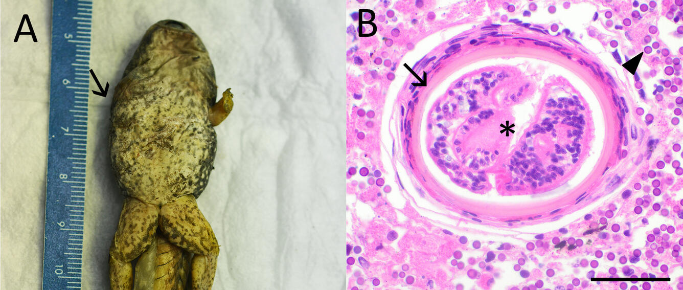
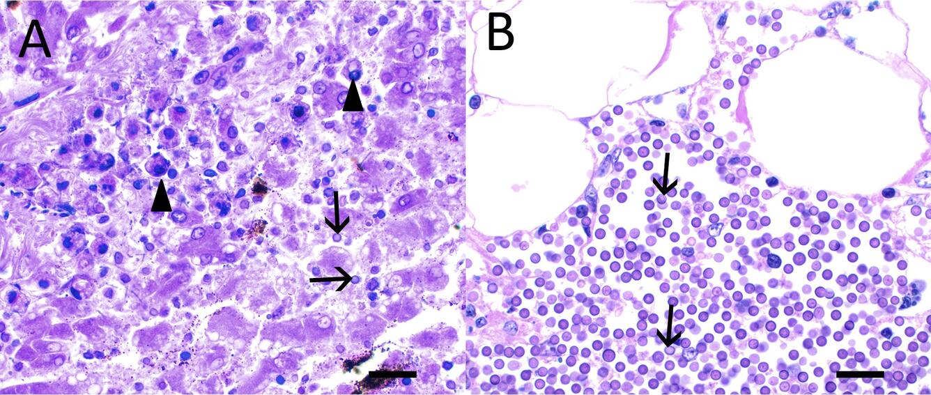
Histopathological Findings: In tadpoles, there are myriad, multifocal to massively extensive sheets and clusters of extra- and intra-cellular, round, 4-6 µm protozoal organisms with a basophilic outer ring and a homogenous amphiphilic interior (Figs. 1B and 2B). These efface the liver, kidneys, and pancreas in many of the tadpoles, are also found in muscle, fat bodies, bone, and in one instance retina and meninges, and are associated with necrosis of host tissues and mild to absent inflammation. In the frog, significantly fewer Perkinsea are present and there is a florid inflammatory response in liver (Fig 2a), kidney, and skin. Occasional encysted trematodes measuring 120 µm in diameter with a thick, hyaline wall (Fig. 1B) are present within necrotic skeletal muscle of tadpoles.
Gross and Morphologic Diagnosis:
- Tadpoles. Extensive, severe, multisystem necrosis with effacement of multiple tissues by myriad Perkinsea-like organisms.
- Frog. Marked granulocytic and granulomatous necrotizing hepatitis, nephritis, and dermatitis with intralesional Perkinsea-like organisms.
- Tadpole. Amelia of the right forelimb, associated with encysted trematode metacercaria.
Disease: Severe Perkinsosis
Etiology: Severe Perkinsea infections are associated with a specific group of Perkinsea protists termed Pathogenic Perkinsea Clade (PPC). Other, similar Perkinsea species have been seen in many species of tadpoles and with a global distribution, but not causing mass mortalities.
Amelia in frogs has been attributed to the presence of toxins, UV light, and infection of the developing limb bud with trematode metacercaria.
Distribution: Subtropical, temperate, and boreal North America. Most outbreaks have been detected along the Gulf of Mexico and Atlantic Coast, but have also occurred in Minnesota, Wisconsin, Alaska, and Oregon. Affected areas often have recurrent outbreaks.
Seasonality: In warmer areas of the east and gulf coasts, outbreaks occur year-round. In cooler temperate regions they have been reported from June to November.
Host range: Severe Perkinsea infections have been reported most often in Lithobates spp. (e.g., green frogs, gopher frogs, leopard frogs, and bullfrogs), including the endangered dusky gopher frog (L. servosa), but are also reported in spring peepers (Pseudacris crucifer), southern cricket frogs (Acris gryllus), and southern leopard frogs (Rana sphenocephalia). Infection has also been seen in a captive population of European tree frogs (Hyla arborea) in the United Kingdom. Prevalence and intensity of infection are much higher in tadpoles compared to adults, and adults are rarely impacted during mass mortalities.
Transmission: Transmission is unknown, but infection is speculated to occur by ingestion of zoospores within the water column. Spores are hardy in the environment, so affected areas are subject to recurrent mortality events.
Clinical signs: During mass mortality events, the most affected life stages are larval, Gosner stages 26-41, which may be sluggish, swimming erratically, circling, or found dead. External lesions are often absent, but abdominal distension, erythema, and petechial hemorrhages on the skin may be present. Dead individuals are often in good to excellent body condition.
Pathology: Gross lesions may include hepatomegaly and pallor, splenomegaly and/or nephromegaly, and thickened intestines. Affected organs may be reddened or pale, with petechial hemorrhages. Ascites or edema may be present. Histologic lesions consist of multifocal clusters to extensive sheets of myriad, 4-6 μm, intra- and extra-cellular protozoal spores with a deep basophilic capsule and eosinophilic to amphiphilic interior, surrounded by necrotic debris and fibrin. These are most often within liver, kidney, and pancreas, but any tissue may be affected. Smaller, 2 μm zoospores lacking the capsule may also be present. Perkinsea organisms efface and destroy large areas of tissue, and inflammation is typically minimal to absent.
Diagnosis: Diagnosis is through histology and PCR.
Public health concerns: None.
Wildlife population impacts: Severe Perkinsea infection is responsible for mass mortalities, causes high mortality (up to 95%) of tadpoles in affected areas, and is a contributor to amphibian decline as the third most common disease of frogs in the United States. This has the potential to drive already imperiled species to extinction.
Management: Removing dead tadpoles from an affected area may reduce environmental pathogen loads.
References:
Chambouvet A, Glower D, Jirků M, Yabsley J, Davis AK, Leonard G, Maguire F, Doherty-Bone TM, Bittencourt-Silva GB, Wilkinson M, Richards TA. 2015. Cryptic infection of a broad taxonomic and geographic diversity of tadpoles by Perkinsea protists. PNAS 112:34. https://doi.org/10.1073/pnas.1500163112
Christiansen J and Feltman H. 2000. A relationship between trematode metacercaria and bullfrog limb abnormalities. Jour. Iowa. Acad. Sci. 107:3. https://scholarworks.uni.edu/jias/vol107/iss3/8
Isodoro-Ayaza M, Lorch JM, Grear DA, Winzler M, Calhoun DL, Barichivich WJ. 2017. Pathogenic lineage of Perkinsea associated with mass mortality of frogs across the United States. Sci. Rep. 7:10288. https://doi.org/10.1038/s41598-017-10456-1
Isodoro-Ayaza M and Grear D. 2019. Pathology and case definition of severe Perkinsea infection of frogs. Vet Path 56:1. https://doi.org/10.1177/0300985818798132
Karwacki EE, Atkinson MS, Ossiboff RJ, Savage AE. 2018. Novel quantitative PCR assay specific for the emerging Perkinsea amphibian pathogen reveals seasonal infection dynamics. Di.s Aquat. Org. 129:85-98. https://doi.org/10.3354/dao03239
Smilansky V, Jirků M, Milner D, Ibáñez R, Gratwicke B, Nichols A, Lukeš J, Chambouvet A, Richards TA. 2021. Extended host and geographic range of tadpole associations with Severe Perkinsea Infection group. Biol. Lett. 17:20210166. https://doi.org/10.1098/rsbl.2021.0166
Related

WHISPers

Pathology Case of the Month

Diagnostic Services
Related
WHISPers
Pathology Case of the Month