
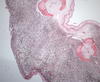
Photomicrograph Showing Melanoma on a Fin of a Brown Bullhead
By Environmental Health Program
2016 (approx.)

Detailed Description
Photomicrograph showing a melanoma on a fin of a brown bullhead fish. In the upper right-hand part is normal skin (epidermis). The epidermis has large clear cells and a layer of normal melanocytes along the basement membrane of the epidermis which is where they normally are located. Most of the rest of the tissue is composed of neoplastic melanocytes that invade up into the epidermis as well as into the dermal and subdermal areas.
Sources/Usage
Public Domain.
Related

Prevalence of Malignant Melanoma in Brown Bullhead from Lake Memphremagog Greater than Expected—Linkages to Contaminant Exposure and Implications for Fish Population Health are Currently Unknown
Raised black lesions observed in 30 percent of the brown bullhead collected from two sites in Lake Memphremagog from 2014 through 2017 were identified microscopically as malignant melanoma. Malignant melanoma in freshwater fishes has been reported before, but this cancer occurrence cluster is raising questions about the cause of the tumors and the implications for the long-term health of fish...
Related
Prevalence of Malignant Melanoma in Brown Bullhead from Lake Memphremagog Greater than Expected—Linkages to Contaminant Exposure and Implications for Fish Population Health are Currently Unknown
Raised black lesions observed in 30 percent of the brown bullhead collected from two sites in Lake Memphremagog from 2014 through 2017 were identified microscopically as malignant melanoma. Malignant melanoma in freshwater fishes has been reported before, but this cancer occurrence cluster is raising questions about the cause of the tumors and the implications for the long-term health of fish...