U.S. flag

An official website of the United States government

Images

Images from the National Wildlife Health Center.

Filter Total Items: 235
Plants growing in lab flasks under lights.
Plants grown to investigate the role of prion contaminated plants in CWD transmission dynamics

Plants were grown in prion-spiked growth media at the National Wildlife Health Center to investigate the role of prion contaminated plants in CWD transmission dynamics.

NWHC pathologist performs skin biopsy from North American racer

National Wildlife Health Center pathologist Susan Knowles obtains a skin biopsy for analysis under a microscope from a Coluber constrictor (North American racer) submitted for diagnostic testing.

National Wildlife Health Center pathologist Susan Knowles obtains a skin biopsy for analysis under a microscope from a Coluber constrictor (North American racer) submitted for diagnostic testing.

Images of heart, pancreas, lung, and brain tissue from gyrfalcon.
Images of heart, pancreas, lung, and brain tissue from gyrfalcon

A. Heart, multifocal and focally extensive necrotizing myocarditis. H&E. Inset: Section of same heart tissue; immunohistochemical staining of influenza A virus antigen is strongly positive in areas of necrosis. B. Pancreas, multifocal and focally extensive necrosis of the acinar epithelium. H&E.

A. Heart, multifocal and focally extensive necrotizing myocarditis. H&E. Inset: Section of same heart tissue; immunohistochemical staining of influenza A virus antigen is strongly positive in areas of necrosis. B. Pancreas, multifocal and focally extensive necrosis of the acinar epithelium. H&E.

NWHC Necropsy
NWHC Necropsy
NWHC Necropsy
NWHC Necropsy

Necropsy of bird performed by NWHC pathologist. 

Necropsy of bird performed by NWHC pathologist. 

Image: UV Light Showing White-Nose Syndrome in Bat's Wing
UV Light Showing White-Nose Syndrome in Bat's Wing

Long-wave ultraviolet (UV) and white-light illumination of lesions associated with white-nose syndrome.  Wing from dead Tri-colored bat (Perimyotis subflavus) lit from above with hand-held 51 LED 385-nm UV flashlight shows points of orange–yellow fluorescence.  

Long-wave ultraviolet (UV) and white-light illumination of lesions associated with white-nose syndrome.  Wing from dead Tri-colored bat (Perimyotis subflavus) lit from above with hand-held 51 LED 385-nm UV flashlight shows points of orange–yellow fluorescence.  

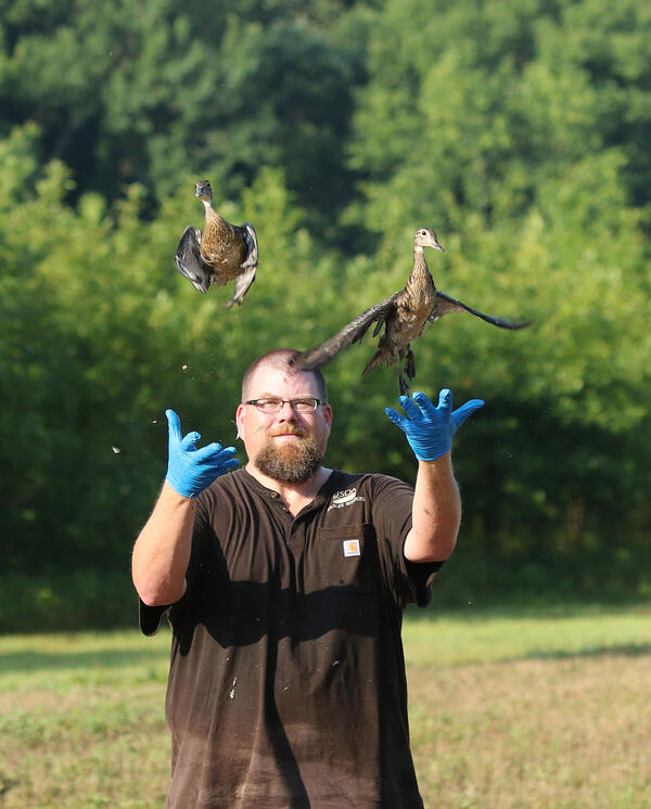
U.S. Department of Agriculture scientist releases a wood duck (Aix sponsa) after collecting a sample for avian influenza
Clint Turnge and wood ducks.

2016 NWRC Accomplishment Report Photos. In 2016, NWRC's National Wildlife Disease Program sampled more than 21,800 wild birds for highly pathogenic avian influenza (HPAI).

2016 NWRC Accomplishment Report Photos. In 2016, NWRC's National Wildlife Disease Program sampled more than 21,800 wild birds for highly pathogenic avian influenza (HPAI).

Pacific Coast
Pacific Coast
Pacific Coast
A moist swab is passed over the surface of the forearm and muzzle of each bat.
Bat being swabbed to test for presence of Pd DNA

A moist swab is passed over the surface of the forearm and muzzle of each bat. The swab will be analyzed in the lab for the presence of Pseudogymnoascus destructans DNA. 

A moist swab is passed over the surface of the forearm and muzzle of each bat. The swab will be analyzed in the lab for the presence of Pseudogymnoascus destructans DNA. 

Photograph of red fox (Vulpes vulpes) in a prairie
Red fox (Vulpes vulpes) in prairie

A red fox (Vulpes vulpes) at the USGS National Wildlife Health Center prairie. 

Sanderling on sand.
Sanderling (Calidris alba)

Sanderling (Calidris alba) walking on sand, seen during field work in Iceland in 2014.

Sanderling (Calidris alba) walking on sand, seen during field work in Iceland in 2014.

Northern gannet flying with mountains in background.
Northern gannet (Morus bassanus)

Northern gannet (Morus bassanus) seen during field work in Iceland in 2013.     

Northern gannet flying with mountains in background.
Northern gannet (Morus bassanus)
Lesser black-backed gull

Lesser black-backed gull flying over water. 

Milk snake (Lampropeltis triangulum) showing signs of fungal and bacterial infections.
Milk snake showing signs of fungal and bacterial infections

Milk snake (Lampropeltis triangulum) showing signs of fungal and bacterial infections, captured in Westchester County, New York, February 2013 (case 24281).

Milk snake (Lampropeltis triangulum) showing signs of fungal and bacterial infections, captured in Westchester County, New York, February 2013 (case 24281).

Pygmy rattlesnake with raised lumps and roughened and crusty scales

Pygmy rattlesnake (Sistrurus miliarius) with multiple raised lumps (nodules) in the dorsal skin of the lower body and tail covered with roughened and crusty scales, captured in Volusia County, Florida, in October 2012.

Pygmy rattlesnake (Sistrurus miliarius) with multiple raised lumps (nodules) in the dorsal skin of the lower body and tail covered with roughened and crusty scales, captured in Volusia County, Florida, in October 2012.

Coral Sampling NWHC HFS
Coral Sampling by the NWHC Honolulu Field Station

Coral Sampling by the National Wildlife Health Center Honolulu Field Station team. 

Was this page helpful?