U.S. flag

An official website of the United States government

Images

Images from the National Wildlife Health Center.

Filter Total Items: 235
Photomicrographs from a big brown bat nasal cavity and hair follicle.
Photomicrographs from a big brown bat (Eptesicus fuscus) from Wisconsi

Photomicrographs from a big brown bat (Eptesicus fuscus) from Wisconsin, USA. H&E stain. (A) Low magnification of a cross section through the nares showing nasal cavity (*), oral mucosa (arrowhead), and haired skin (arrow). (B) Higher magnification showing a normal hair follicle (arrow) surrounded by sebaceous glands.

Photomicrographs from a big brown bat (Eptesicus fuscus) from Wisconsin, USA. H&E stain. (A) Low magnification of a cross section through the nares showing nasal cavity (*), oral mucosa (arrowhead), and haired skin (arrow). (B) Higher magnification showing a normal hair follicle (arrow) surrounded by sebaceous glands.

Photographs of mallard duck lungs with arrows pointing to white to tan nodules.
Photographs of lungs from a mallard duck found dead in Idaho

Photographs from a mallard duck (Anas platyrhynchos) found dead in Idaho, USA.  (A) The lungs are dark red and have multifocal 1-3 mm diameter white to tan nodules disseminated throughout (arrows).  (B) Cut section of the lung showing the nodules within the parenchyma (arrows).

Photographs from a mallard duck (Anas platyrhynchos) found dead in Idaho, USA.  (A) The lungs are dark red and have multifocal 1-3 mm diameter white to tan nodules disseminated throughout (arrows).  (B) Cut section of the lung showing the nodules within the parenchyma (arrows).

Photomicrographs of lung from a mallard duck showing fungal hyphae.
Photomicrographs of lung from a mallard duck

Photomicrographs from a mallard duck (Anas platyrhynchos) found dead in Idaho, USA.  (A) Multiple granulomas (*) with eosinophilic necrotic centers efface the lung. H&E stain.  (B) Granulomas contain many degenerate heterophils and a few multinucleated giant cells (arrows). H&E stain.

Photomicrographs from a mallard duck (Anas platyrhynchos) found dead in Idaho, USA.  (A) Multiple granulomas (*) with eosinophilic necrotic centers efface the lung. H&E stain.  (B) Granulomas contain many degenerate heterophils and a few multinucleated giant cells (arrows). H&E stain.

Gloved hands holding a bat white glove scientists administers vaccine orally.
Bat receiving white-nose syndrome vaccine

A bat receiving the white-nose syndrome vaccine during a field trial to study vaccine efficacy. 

Four photographs from a red wolf including heartworms in heart and abnormalities on legs.
Photographs from a red wolf (Canis rufus) found dead in North Carolina

Photographs from a red wolf (Canis rufus) found dead in North Carolina, USA. (A) Adult heartworms (Dirolfilaria immitis) in the right ventricle and atria of the heart and extending into the pulmonary artery. (B) Hard, haired nodule on the medial surface of the distal radius of the right leg.

Photographs from a red wolf (Canis rufus) found dead in North Carolina, USA. (A) Adult heartworms (Dirolfilaria immitis) in the right ventricle and atria of the heart and extending into the pulmonary artery. (B) Hard, haired nodule on the medial surface of the distal radius of the right leg.

Histology panel from eastern gray squirrel

Photomicrographs from the lung of an eastern gray squirrel (Sciurus carolinensis) found dead in Wisconsin, U.S.A. (A) A bronchiole (arrow) contains numerous neutrophils. H&E stain. Inset: Bronchiolar epithelium is overlain by Gram-negative bacteria. Brown and Hopps stain. Bar = 20 µm.

Photomicrographs from the lung of an eastern gray squirrel (Sciurus carolinensis) found dead in Wisconsin, U.S.A. (A) A bronchiole (arrow) contains numerous neutrophils. H&E stain. Inset: Bronchiolar epithelium is overlain by Gram-negative bacteria. Brown and Hopps stain. Bar = 20 µm.

Necropsy photos from eastern gray squirrel

Photographs from an eastern gray squirrel (Sciurus carolinensis) found dead in Wisconsin, U.S.A.  (A) The lung contained multifocal firm red areas (arrows) and multifocal areas of hemorrhage (arrowhead). (B) Firm areas are red to tan on cut section and airways contain mucoid red to tan fluid.

Photographs from an eastern gray squirrel (Sciurus carolinensis) found dead in Wisconsin, U.S.A.  (A) The lung contained multifocal firm red areas (arrows) and multifocal areas of hemorrhage (arrowhead). (B) Firm areas are red to tan on cut section and airways contain mucoid red to tan fluid.

Photomicrographs from a desert cottontail showing changes due to RHDV2.
Photomicrographs from a desert cottontail found dead in Texas

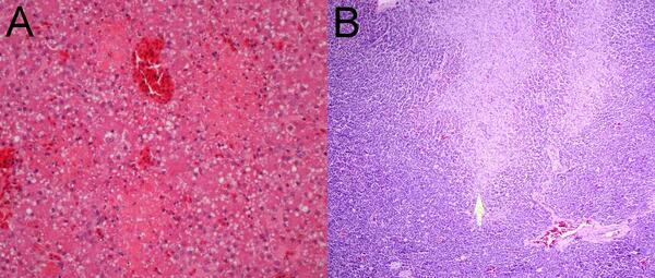
Photomicrographs from a desert cottontail found dead in Texas, U.S.A. showing (A) panlobular hepatocellular dissociation and necrosis and (B) intra-alveolar edema fluid (asterisk) and foamy pulmonary macrophages (arrows) in the lung.

Photomicrographs from a desert cottontail found dead in Texas, U.S.A. showing (A) panlobular hepatocellular dissociation and necrosis and (B) intra-alveolar edema fluid (asterisk) and foamy pulmonary macrophages (arrows) in the lung.

Microscopic view of organs of mountain cottontail rabbit.
Photographs from organs of a mountain cottontail rabbit found dead

Photographs from an adult female mountain cottontail rabbit (Sylvilagus nuttallii) found dead in Montana, U.S.A. (A) Liver with random foci of necrosis (asterisk) characterized by accumulation of cellular detritus intermingled with fibrin and colonies of small coccoid bacteria (inset). H&E stain.

Photographs from an adult female mountain cottontail rabbit (Sylvilagus nuttallii) found dead in Montana, U.S.A. (A) Liver with random foci of necrosis (asterisk) characterized by accumulation of cellular detritus intermingled with fibrin and colonies of small coccoid bacteria (inset). H&E stain.

Photographs from mallard ducks showing trematodes in tissue.
Photographs from two mallards (Anas platyrhynchos) with trematodiasis

Photographs from two mallards (Anas platyrhynchos) found dead within a warm-water retention pond in Minnesota, US. (A) Jejunum with superficial pseudomembranes and deep mucosal granulomatous inflammation (clear arrow). Small degenerated metazoan parasites are present rarely within the inflamed areas (black arrow). H&E stain. (B).

Photographs from two mallards (Anas platyrhynchos) found dead within a warm-water retention pond in Minnesota, US. (A) Jejunum with superficial pseudomembranes and deep mucosal granulomatous inflammation (clear arrow). Small degenerated metazoan parasites are present rarely within the inflamed areas (black arrow). H&E stain. (B).

Photographs of tissues samples from mallard duck intestine next to ruler for scale.
Photographs of intestine tissue from mallard ducks with trematodiasis

Photographs from two mallards (Anas platyrhynchos) found dead within a warm-water retention pond in Minnesota, US. (A) Caeca with mucosal roughening and caseous plaque formation. A larger trematode (arrow) is found within. (B) Dilated and thinned intestine containing watery intestinal content with pinpoint white material.

Photographs from two mallards (Anas platyrhynchos) found dead within a warm-water retention pond in Minnesota, US. (A) Caeca with mucosal roughening and caseous plaque formation. A larger trematode (arrow) is found within. (B) Dilated and thinned intestine containing watery intestinal content with pinpoint white material.

Photomicrographs from a Common Eider showing necrosis.
Photomicrographs from a Common Eider (Somateria mollissima)

Photomicrographs from a Common Eider (Somateria mollissima) from Massachusetts, USA. (A) Multiple pale foci of acute hepatic necrosis. (B) Multiple foci of acute pancreatic necrosis (arrow).

Photomicrographs from a Common Eider (Somateria mollissima) from Massachusetts, USA. (A) Multiple pale foci of acute hepatic necrosis. (B) Multiple foci of acute pancreatic necrosis (arrow).

Photographs of a common eider liver and pancreas.
Photographs from a Common Eider (Somateria mollissima)

Photographs from a Common Eider (Somateria mollissima) from Massachusetts, USA. (A) There are scattered, small, pale foci in the liver. (B) Petechial and ecchymotic hemorrhages are present in the pancreas.

Photographs from a Common Eider (Somateria mollissima) from Massachusetts, USA. (A) There are scattered, small, pale foci in the liver. (B) Petechial and ecchymotic hemorrhages are present in the pancreas.

A pathologist performs a necropsy on a rabbit
Pathologist examines a cottontail for cause-of-death determination
Grey colored Cope’s grey treefrog camouflaged against a tree trunk.
Cope’s grey treefrog

Cope’s grey treefrog on a tree trunk. Seen during fieldwork in Wisconsin.

Cope’s grey treefrog on a tree trunk. Seen during fieldwork in Wisconsin.

View of wetland vegetation with a camouflaged green frog.
Green frog in wetland vegetation

A green frog camouflaged in wetland vegetation that was seen during fieldwork in Wisconsin.

A green frog camouflaged in wetland vegetation that was seen during fieldwork in Wisconsin.

View of ground covered in leaf litter with a camouflaged American toad.
American toad tucking into the leaf litter

American toad tucking into the leaf litter seen during fieldwork in Wisconsin.

Green colored Cope’s grey treefrog on a tree trunk.
Cope’s grey treefrog, green morph

Cope’s grey treefrog, green morph, on a tree trunk. Seen during fieldwork in Wisconsin.

Was this page helpful?