
Scientists at the USGS National Wildlife Health Center have been working on vaccines to protect bats from diseases like such as rabies and white-nose syndrome. The most promising method is called orotopical vaccination. By applying the vaccine to the bat’s muzzle, the gel-based vaccine is spread to others in the colony through grooming.
Images
Images from the National Wildlife Health Center.
Filter Total Items: 229
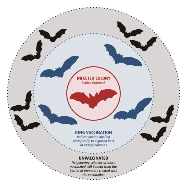
Ring Vaccination - Rabies and Vampire Bats
Scientists at the USGS National Wildlife Health Center have been working on vaccines to protect bats from diseases like such as rabies and white-nose syndrome. The most promising method is called orotopical vaccination. By applying the vaccine to the bat’s muzzle, the gel-based vaccine is spread to others in the colony through grooming.
photograph by Anna Pink Snowy owl
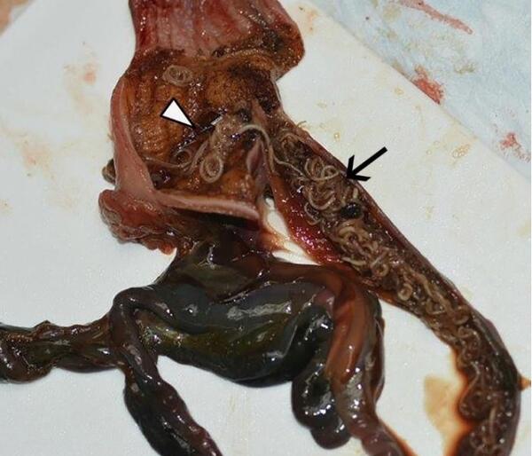
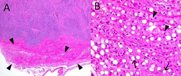
Photograph from a Snowy Owl (Bubo scandiacus) found dead in Wisconsin, USA. Numerous nematodes are present in the ventriculus (white arrowhead) and duodenum (black arrow).
Photograph from a Snowy Owl (Bubo scandiacus) found dead in Wisconsin, USA. Numerous nematodes are present in the ventriculus (white arrowhead) and duodenum (black arrow).
Scientists investigate pathogens in salamanders
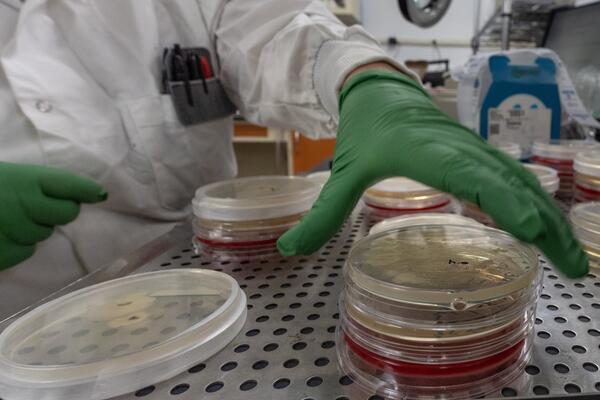
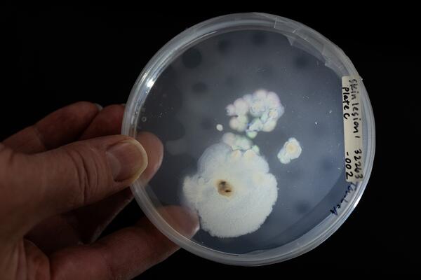
A USGS scientist reaches for a microbiological plate with cultures from salamander skin to investigate for potential pathogens.
A USGS scientist reaches for a microbiological plate with cultures from salamander skin to investigate for potential pathogens.
Scientists investigate pathogens in salamanders
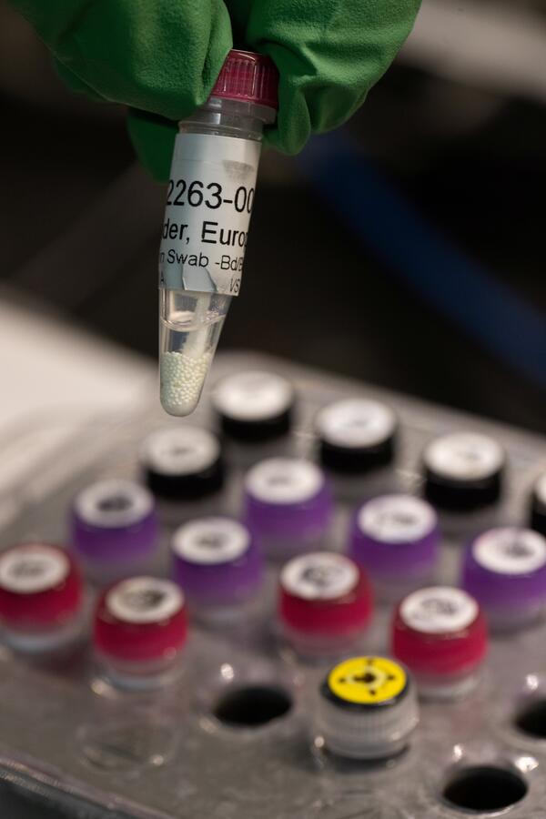
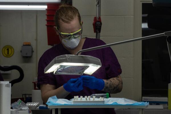
A USGS scientist prepares to analyze a skin swab from a salamander to investigate for potential pathogens.
A USGS scientist prepares to analyze a skin swab from a salamander to investigate for potential pathogens.
Scientists investigate pathogens in salamanders
A USGS scientist examines a skin sample from a salamander as part of a study to investigate for potential pathogens.
A USGS scientist examines a skin sample from a salamander as part of a study to investigate for potential pathogens.
Scientists investigate pathogens in salamanders
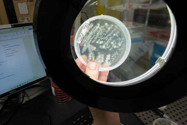
A USGS scientist examines microorganisms from salamander skin to investigate for potential pathogens.
A USGS scientist examines microorganisms from salamander skin to investigate for potential pathogens.
Holiday hours banner
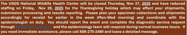
Thanksgiving Banner_ Holiday hours banner "The USGS National Wildlife Health Center will be closed Thursday, Nov 27, 2025 and have reduced staffing on Friday, Nov 28, 2025 for the Thanksgiving holiday which may affect your shipments, submission processing and results reporting. Please plan your specimen collections and shipments accordingly for r
Thanksgiving Banner_ Holiday hours banner "The USGS National Wildlife Health Center will be closed Thursday, Nov 27, 2025 and have reduced staffing on Friday, Nov 28, 2025 for the Thanksgiving holiday which may affect your shipments, submission processing and results reporting. Please plan your specimen collections and shipments accordingly for r
Scientists investigate pathogens in salamanders
A USGS scientist examines salamanders as part of a study to investigate for potential pathogens.
A USGS scientist examines salamanders as part of a study to investigate for potential pathogens.
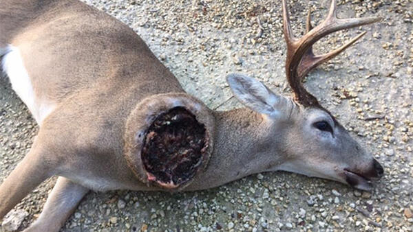
New World Screwworm On an Open Wound
New World Screwworm is a fly (Cochliomyia hominivorax) that lays eggs in open wounds of warm-blooded animals. Once hatched, the larvae (maggots) consume the living tissues at the edge of the wound, leading to severe illness and death if left untreated.
New World Screwworm is a fly (Cochliomyia hominivorax) that lays eggs in open wounds of warm-blooded animals. Once hatched, the larvae (maggots) consume the living tissues at the edge of the wound, leading to severe illness and death if left untreated.
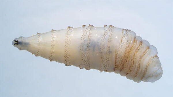
New World Screwworm larvae
New World Screwworm is a fly (Cochliomyia hominivorax) that lays eggs in open wounds of warm-blooded animals. Once hatched, the larvae (maggots) consume the living tissues at the edge of the wound, leading to severe illness and death if left untreated.
New World Screwworm is a fly (Cochliomyia hominivorax) that lays eggs in open wounds of warm-blooded animals. Once hatched, the larvae (maggots) consume the living tissues at the edge of the wound, leading to severe illness and death if left untreated.
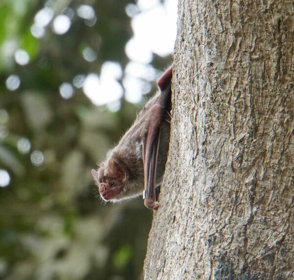
Vampire Bat on a Tree
USGS is developing and testing a safe rabies vaccine for vampire bats and conducting field studies for a practical delivery of this vaccine to wild populations of bats in the future.
USGS is developing and testing a safe rabies vaccine for vampire bats and conducting field studies for a practical delivery of this vaccine to wild populations of bats in the future.
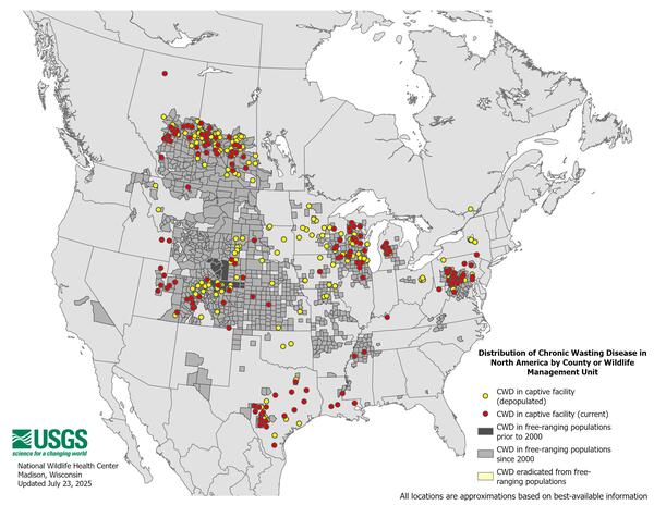
Distribution of Chronic Wasting Disease in North America from 2000 through July 2025.
Distribution of Chronic Wasting Disease in North America from 2000 through July 2025.Distribution of Chronic Wasting Disease in North America, updated July 23, 2025.
Distribution of Chronic Wasting Disease in North America from 2000 through July 2025.
Distribution of Chronic Wasting Disease in North America from 2000 through July 2025.Distribution of Chronic Wasting Disease in North America, updated July 23, 2025.
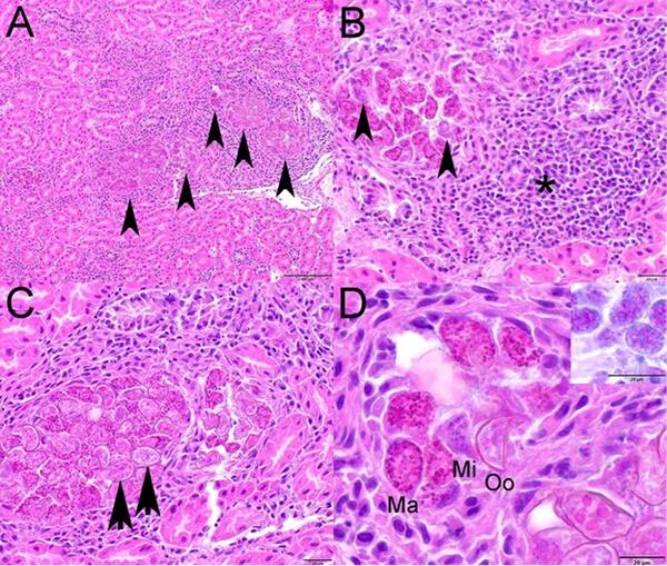
Photomicrographs from a Canada Goose (Branta canadensis) from Ohio, USA.
Photomicrographs from a Canada Goose (Branta canadensis) from Ohio, USA.Photomicrographs from a Canada Goose (Branta canadensis) from Ohio, USA. Hematoxylin and eosin stain. (A) Renal tubules are distended with various stages of intraepithelial coccidia (arrowheads) and are surrounded by inflammation. (B) Macrogamonts and fewer microgamonts (arrowheads) expa
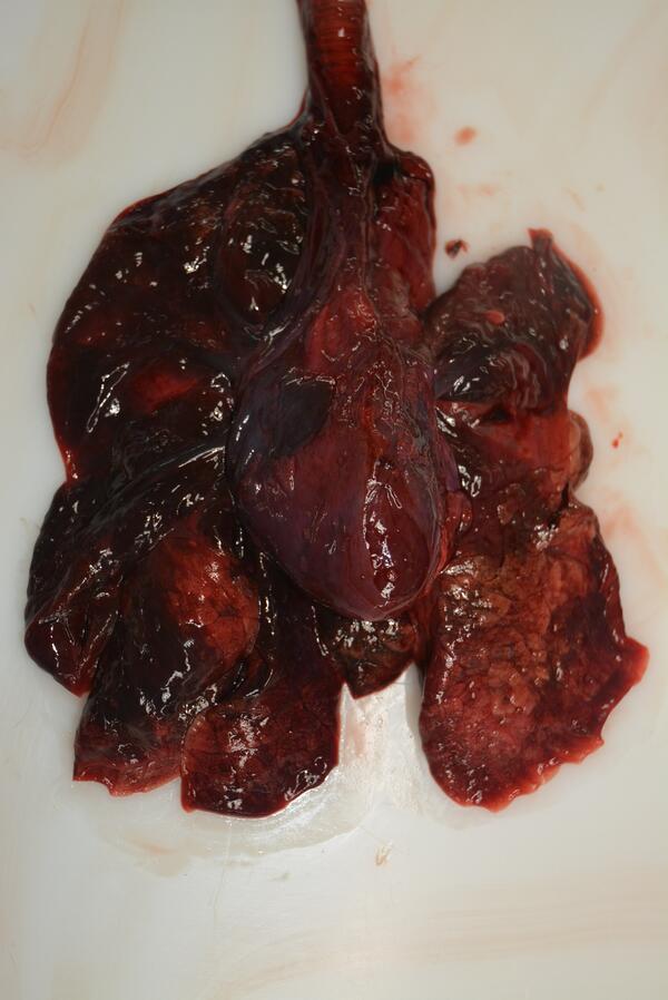
Northern Sea Otter May 2025-01
Emaciation with serous atrophy of fat in a Northern Sea Otter Case Of The Month 2025
Emaciation with serous atrophy of fat in a Northern Sea Otter Case Of The Month 2025
Emaciation with serous atrophy of fat in a Northern Sea Otter (Enhydra lutris)
Emaciation with serous atrophy of fat in a Northern Sea Otter (Enhydra lutris)Figure 1. Photomicrographs from a Northern Sea Otter (Enhydra lutris) found dead on a beach in Alaska, USA. (A) Lymph node. Germinal centers are depleted. Adjacent mesenteric adipose tissue markedly atrophic (arrowheads). (B) High magnification of mesenteric adipose tissue.

Distribution of CWD in Relation to Tribal Lands in the U.S.
Distribution of CWD in Relation to Tribal Lands in the U.S.Distribution of Chronic Wasting Disease in North America, in Relation to Tribal Lands in the Conterminous United States. Updated on April 17, 2025.
Distribution of CWD in Relation to Tribal Lands in the U.S.
Distribution of CWD in Relation to Tribal Lands in the U.S.Distribution of Chronic Wasting Disease in North America, in Relation to Tribal Lands in the Conterminous United States. Updated on April 17, 2025.

Distribution of Chronic Wasting Disease in North America
Distribution of Chronic Wasting Disease in North AmericaDistribution of Chronic Wasting Disease in North America, updated April 11, 2025.
Distribution of Chronic Wasting Disease in North America
Distribution of Chronic Wasting Disease in North AmericaDistribution of Chronic Wasting Disease in North America, updated April 11, 2025.

Chronic wasting disease: change in documented distribution in North America 2000-2024
Chronic wasting disease: change in documented distribution in North America 2000-2024Animated GIF showing changes in distribution of chronic wasting disease in North America from 2000-2024 as documented at the end of each year. Individual files of yearly maps are available at https://doi.org/10.5066/P9HQKKFO.

Distribution of Highly Pathogenic Avian Influenza H5 and H5N1 in North America, 2021-2025
Distribution of Highly Pathogenic Avian Influenza H5 and H5N1 in North America, 2021-2025Distribution of Highly Pathogenic Avian Influenza H5 and H5N1 in wild birds the United States, by county, 2021-2025. Updated February 10, 2025.

Dr. LeAnn White has been selected as the new Center Director of the USGS National Wildlife Health Center (NWHC)
Dr. LeAnn White has been selected as the new Center Director of the USGS National Wildlife Health Center (NWHC)Dr. LeAnn White has been selected as the new Center Director of the USGS National Wildlife Health Center (NWHC)

Conceptual illustration of the future USGS National Wildlife Health Center, courtesy of Flad Architects
Conceptual illustration of the future USGS National Wildlife Health Center, courtesy of Flad ArchitectsNWHC modernization overhead view, The image showcases an overhead view of the NWHC modernization project for the microsite homepage. Its highlighting the contemporary structures integrated into a vibrant landscape. Provided by: Lankton, Julia