
The front elevation showcases a combination of glass and metal elements, reflecting a contemporary aesthetic. WHC will be a single, integrated, multistory, state-of-the-art building that will include offices, laboratories (BSL-2 and BSL-3), and vivarium
Picture provide by :Julia Lankton
Deputy Center Director
Images
Images from the National Wildlife Health Center.
Filter Total Items: 229

NWHC modernization
The front elevation showcases a combination of glass and metal elements, reflecting a contemporary aesthetic. WHC will be a single, integrated, multistory, state-of-the-art building that will include offices, laboratories (BSL-2 and BSL-3), and vivarium
Picture provide by :Julia Lankton
Deputy Center Director
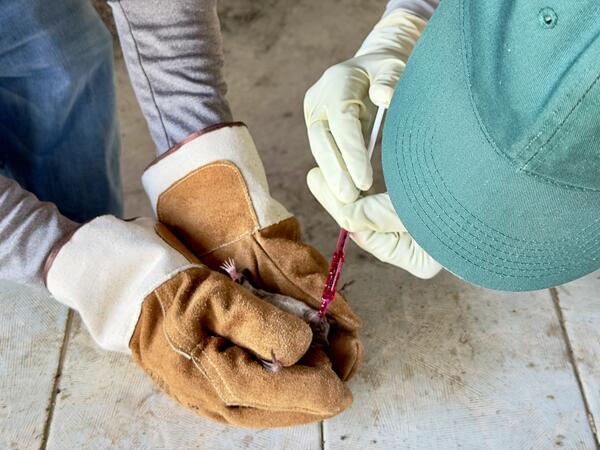
Applying Vaccine to a Vampire Bat
USGS is developing and testing a safe rabies vaccine for vampire bats and conducting field studies for a practical delivery of this vaccine to wild populations of bats in the future.
USGS is developing and testing a safe rabies vaccine for vampire bats and conducting field studies for a practical delivery of this vaccine to wild populations of bats in the future.

Photographs from a mountainous star coral (Montastraea cavernosa) losing tissue to disease in Florida, USA.
Photographs from a mountainous star coral (Montastraea cavernosa) losing tissue to disease in Florida, USA.Photographs from a mountainous star coral (Montastraea cavernosa) losing tissue to disease in Florida, USA. (A) Colony in situ showing bare white, recently denuded skeleton with no turf algae growth (arrowheads) with dark discoloration along the active tissue loss margins (arrows).

Photomicrographs from a mountainous star coral (Montastraea cavernosa) in Florida, USA.
Photomicrographs from a mountainous star coral (Montastraea cavernosa) in Florida, USA.Photomicrographs from a mountainous star coral (Montastraea cavernosa) in Florida, USA. (A) A large cluster of filamentous bacteria are within the gastrovascular space and gastrodermis (asterisk) and extending through mesoglea (arrow) with necrosis and loss of the surface gastrodermis (arrowheads). H&E stain. Scale bar 200 μm.

Photomicrographs from a sea otter (Enhydra lutris) found dead in Washington State, USA.
Photomicrographs from a sea otter (Enhydra lutris) found dead in Washington State, USA.Photomicrographs from a sea otter (Enhydra lutris) found dead in Washington State, USA.
Photomicrographs from a sea otter (Enhydra lutris) found dead in Washington State, USA.
Photomicrographs from a sea otter (Enhydra lutris) found dead in Washington State, USA.Photomicrographs from a sea otter (Enhydra lutris) found dead in Washington State, USA.

Photographs from a Little Brown Bat (Myotis lucifugus) found dead in Montana, USA.
Photographs from a Little Brown Bat (Myotis lucifugus) found dead in Montana, USA.Photographs from a Little Brown Bat (Myotis lucifugus) found dead in Montana, USA (A, B, D). (A) Multiple foci of depigmentation (arrowheads) measuring up to 3 mm in diameter are present on the patagia. (B) Under ultraviolet light, there is a single pinpoint focus of orange fluorescence (arrowhead) and multiple pinpoint foci of blue fluorescence (arrow).

Photomicrographs from little brown bats, one with square-eared anomaly, found dead in Montana, USA.
Photomicrographs from little brown bats, one with square-eared anomaly, found dead in Montana, USA.Photomicrographs from an unaffected Little Brown Bat (M. lucifugus; A-B) and Little Brown Bat found dead in Montana, USA with the square-eared anomaly (C-E). Periodic Acid Schiff-Hematoxylin. (A) Pinna of an unaffected Little Brown Bat showing the epidermis (e and arrow), dermis (d) and central elastic cartilage plate (c).
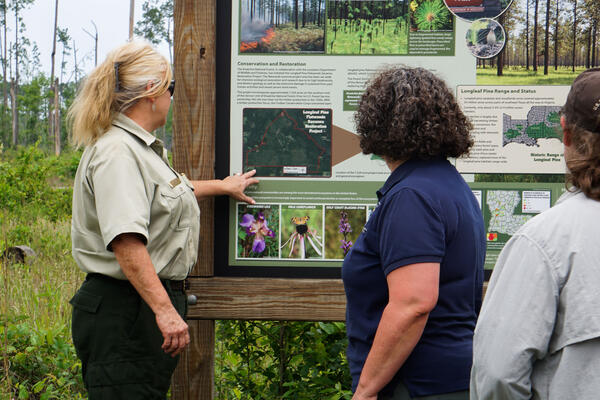
Forest Supervisor talks with USFWS about flatwoods
Forest Supervisor talks with USFWS about flatwoodsKisatchie National Forest Supervisor talks with U.S. Fish and Willdlife Service employees about a flatwoods project site in the forest May 7, 2024.
Forest Supervisor talks with USFWS about flatwoods
Forest Supervisor talks with USFWS about flatwoodsKisatchie National Forest Supervisor talks with U.S. Fish and Willdlife Service employees about a flatwoods project site in the forest May 7, 2024.

Photomicrographs of the small intestine from a Western gull (Larus occidentalis) from California, USA
Photomicrographs of the small intestine from a Western gull (Larus occidentalis) from California, USAPhotomicrographs of the small intestine from a Western gull (Larus occidentalis) from California, USA. H & E stain. (A) Expanding the small intestinal lumen and distorting intestinal villi are multiple adult metazoan parasites (arrow); similar parasites are within cavitations in the intestinal wall or serosa (arrowhead).

Photographs from a Western gull (Larus occidentalis) in California, USA
Photographs from a Western gull (Larus occidentalis) in California, USAPhotographs from a Western gull (Larus occidentalis) in California, USA. (A) On the intestinal serosa are multifocal 1-mm diameter firm white nodules (arrows). A white fungal plaque (white arrow) also overlies the serosa.

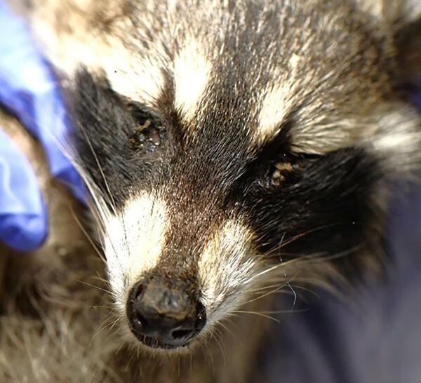
Photographs and photomicrographs from a Common Raccoon (Procyon lotor) found dead in Wisconsin
Photographs and photomicrographs from a Common Raccoon (Procyon lotor) found dead in WisconsinPhotographs and photomicrographs from a Common Raccoon (Procyon lotor) found dead in Wisconsin, USA. (A) There is green-gray mucoid discharge around the eyes and the nasal planum is crusty (arrows).
Racoon001.jpg
Common Raccoon (Procyon lotor) found dead in Wisconsin
Common Raccoon (Procyon lotor) found dead in Wisconsin

Tissue from a gull (Larus sp) from Wisconsin
Tissue from a gull (Larus sp) from Wisconsin. (A) Diffusely the pericardium (star) is greatly expanded by fibrin, edema and necrotic debris (H&E). (B) Lymphoplasmacytic myositis (arrows) creating linear lesions along fascial planes of the pectoral skeletal muscle (H&E).
Tissue from a gull (Larus sp) from Wisconsin. (A) Diffusely the pericardium (star) is greatly expanded by fibrin, edema and necrotic debris (H&E). (B) Lymphoplasmacytic myositis (arrows) creating linear lesions along fascial planes of the pectoral skeletal muscle (H&E).

Photomicrographs of the plagiopatagium from a Little Brown Bat
Photomicrographs of the plagiopatagium from a Little Brown BatPhotomicrographs of the plagiopatagium from a Little Brown Bat (Myotis lucifugus) captured live in Wyoming, U.S.A. (A) Adult nematodes (asterisks) are present in the dermis. They are filled with larval nematodes (arrowhead). The overlying epidermis is hyperplastic (arrow). H&E stain.

Gross photographs from a Cooper’s hawk (Accipiter cooperii)
Gross photographs from a Cooper’s hawk (Accipiter cooperii)Gross photographs from a Cooper’s hawk (Accipiter cooperii). (A) Air sac overlying the lung and liver have multiple yellow to green, irregular nodules (arrows). (B) Liver (asterisk) has multiple 1 mm, round to irregular, tan nodules in addition to innumerable miliary foci.

Photomicrographs of Cooper’s hawk soft palate and lung
Photomicrographs of Cooper’s hawk soft palate and lung(A) Soft palate of a Cooper’s hawk (Accipiter cooperii). The submucosa is expanded by a myriad of epithelioid macrophages (arrow) surrounding multinucleated giant cells centered on a necrotic center (asterisk). H&E stain. Insert: Intrahistiocytic and extracellular acid-fast bacilli.

Photomicrograph from a healthy example of elkhorn coral side-by-side with elkhorn coral found with multifocal tissue loss
Photomicrograph from a healthy example of elkhorn coral side-by-side with elkhorn coral found with multifocal tissue loss
Photomicrograph at low magnification from elkhorn coral (Acropora palmata) found with multifocal tissue loss in the Dominican Republic
Photomicrograph at low magnification from elkhorn coral (Acropora palmata) found with multifocal tissue loss in the Dominican RepublicPhotomicrograph at low magnification from elkhorn coral (Acropora palmata) found with multifocal tissue loss in the Dominican Republic. A flatworm (fw) is present along a multifocally ulcerated coral surface body wall (c) along one polyp. A small amount of cellular debris can be seen within the pharynx (p) of the flatworm.

Photomicrographs of from unknown species of flatworm
Photomicrographs of from unknown species of flatwormPhotomicrographs of from unknown species of flatworm. (A) Dorsal detail showing epidermis with rhabdites (black arrowheads) and endosymbionts visible within the flatworm parenchyma (fw).
Photomicrographs of from unknown species of flatworm
Photomicrographs of from unknown species of flatwormPhotomicrographs of from unknown species of flatworm. (A) Dorsal detail showing epidermis with rhabdites (black arrowheads) and endosymbionts visible within the flatworm parenchyma (fw).

Photographs from an elkhorn coral colony (Acropora palmata) losing tissue on a reef in the Dominican Republic
Photographs from an elkhorn coral colony (Acropora palmata) losing tissue on a reef in the Dominican RepublicPhotographs from an elkhorn coral colony (Acropora palmata) losing tissue on a reef in the Dominican Republic. (A) Colony in situ with bright white skeleton exposed by multifocal tissue loss (arrowheads) and areas of older tissue loss with algae overgrowth (*). (B) Submitted samples. Gross lesions are obscured by with loss of pigment with fixation.

Photographs from a Mexican Wolf (Canis lupus baileyi) euthanized due to neurologic illness in New York
Photographs from a Mexican Wolf (Canis lupus baileyi) euthanized due to neurologic illness in New YorkPhotographs from a Mexican Wolf (Canis lupus baileyi) euthanized due to neurologic illness in New York, USA. (A) An area of light gray discoloration is present on the surface of the left cranio-dorsal cerebrum (arrow).