
Photomicroscopy and Flow Cytometry Core Technology Team
Microscopic Video Imaging
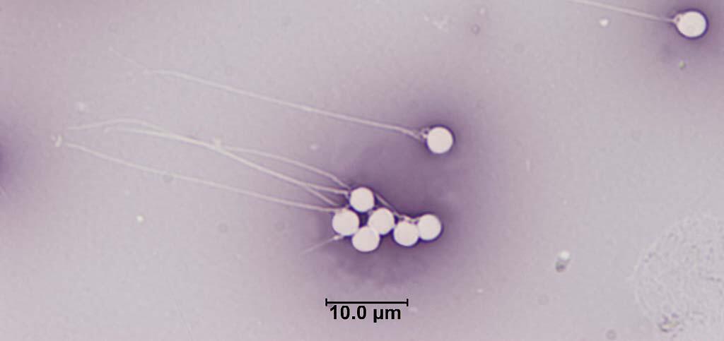
Eosin-nigrosin staining of spermatozoa from common carp
Flow cytometry results from yellow perch testicular tissue
About the Research
The Photomicroscopy and Flow Cytometry Core Technology Team (CTT) as part of the Environmental Health Program works to develop and apply biomarkers to evaluate the potential impacts of environmental contaminants at cellular and molecular levels. Because molecular and biochemical responses of cells are preceded by chemical changes in nuclei, cytoplasm, membranes, and extracellular fluids, these responses can be indicative of contaminant exposures.

Animal biomarkers diagnostic of ecosystem condition: Biotechnologies for conservation science.
The Photomicroscopy and Flow Cytometry CTT focuses on bioindicators associated with environmental stressors and ecosystem integrity, which are essential to the management of trust resources, habitats, and ecosystems that serve human, and fish and wildlife communities.
The CTT uses biotechnologies typically used in human medicine to collect data on animal cells. In general, blood and sperm are used to determine markers of response such as chemical changes in cell structures and DNA, which can indicate animal condition.
Data gathered can reflect reproductive status and indicate genotoxicity (relevant to environmental conditions). This information is often integrated with other organism, population and landscape level information and analytical chemistry data to provide information on adverse outcome pathways (i.e., linkages between a direct molecular initiating event and an adverse outcome at a biological level of organization), as well as biomarkers (e.g., early-warning signals that reflect biological responses). These methods can be used in research with bacteria to mammals to deliver actionable science to support environmental health research and goals.
Key Analytical Capabilities

- Cell viability and motility
- Mitochondrial membrane potential
- Apoptosis (programmed cell death)
- Liver enzymes
- Ploidy
- DNA fragmentation
- Cell cycle/proliferation
- Genome size
- Cell counts
- Blood cell type identification
- Epigenetics
- Immunophenotyping
Key Instrumentation
- Flow Cytometer
- Spectral Flow Cytometry
- Computer Assisted Sperm Motion Analysis (CASA) System
- Microscopes and microscopic image analysis software
Environmental Health Integrated Science Team Collaborators
-
Toxins and Harmful Algal Blooms Science Team
The team develops advanced methods to study factors driving algal toxin production, how and where wildlife or humans are exposed to toxins, and ecotoxicology. That information is used to develop decision tools to understand if toxin exposure leads to adverse health effects in order to protect human and wildlife health.
Per-and Polyfluoroalkyl Substances (PFAS) Integrated Science Team
Increasing scientific and public awareness of the widespread distribution of per- and poly-fluoroalkyl substances (PFAS) in U.S. drinking-water supplies, aquatic and terrestrial ecosystems, wildlife, and humans has raised many public health and resource management questions that U.S. Geological Survey's (USGS) science can inform. The USGS Environmental Health Program's PFAS Integrated Science Team...
Minerals Resources Life Cycle Integrated Science Team
The Minerals Resources Life Cycle Integrated Science Team focuses on contaminant exposures in the environment that might originate from mineral resource activities including, transportation, storage, extraction and waste management. Perceived health risks to humans and other organisms will be distinguished from actual risks, if any. If actual risks are identified the science produced by this team...
Science activities related to the Photomicroscopy and Flow Cytometry Core Technology Team can be found below.
Minerals Resources Life Cycle Integrated Science Team

Flow Cytometry Applied to the Animal Kingdom in Studies of Natural Resource Science

Outlining Potential Health Effects of Exposure to Critical Elements: From Chemical Structure to Adverse Outcome Pathways

Detecting Sublethal Effects of Harmful Algal Blooms in Mammalian and Avian Cells

Comparative Freshwater Fish Toxicity Testing of Antimycin A

Computer-Assisted Sperm Motion Analysis in Measuring Reproductive Effects in Bass

Studying Immune Responses in the American Kestrel (Falco sparverius)
About the Research
The Photomicroscopy and Flow Cytometry Core Technology Team (CTT) as part of the Environmental Health Program works to develop and apply biomarkers to evaluate the potential impacts of environmental contaminants at cellular and molecular levels. Because molecular and biochemical responses of cells are preceded by chemical changes in nuclei, cytoplasm, membranes, and extracellular fluids, these responses can be indicative of contaminant exposures.
Animal biomarkers diagnostic of ecosystem condition: Biotechnologies for conservation science.
The Photomicroscopy and Flow Cytometry CTT focuses on bioindicators associated with environmental stressors and ecosystem integrity, which are essential to the management of trust resources, habitats, and ecosystems that serve human, and fish and wildlife communities.
The CTT uses biotechnologies typically used in human medicine to collect data on animal cells. In general, blood and sperm are used to determine markers of response such as chemical changes in cell structures and DNA, which can indicate animal condition.
Data gathered can reflect reproductive status and indicate genotoxicity (relevant to environmental conditions). This information is often integrated with other organism, population and landscape level information and analytical chemistry data to provide information on adverse outcome pathways (i.e., linkages between a direct molecular initiating event and an adverse outcome at a biological level of organization), as well as biomarkers (e.g., early-warning signals that reflect biological responses). These methods can be used in research with bacteria to mammals to deliver actionable science to support environmental health research and goals.
Key Analytical Capabilities
- Cell viability and motility
- Mitochondrial membrane potential
- Apoptosis (programmed cell death)
- Liver enzymes
- Ploidy
- DNA fragmentation
- Cell cycle/proliferation
- Genome size
- Cell counts
- Blood cell type identification
- Epigenetics
- Immunophenotyping
Key Instrumentation
- Flow Cytometer
- Spectral Flow Cytometry
- Computer Assisted Sperm Motion Analysis (CASA) System
- Microscopes and microscopic image analysis software
Environmental Health Integrated Science Team Collaborators
-
-
Toxins and Harmful Algal Blooms Science Team
The team develops advanced methods to study factors driving algal toxin production, how and where wildlife or humans are exposed to toxins, and ecotoxicology. That information is used to develop decision tools to understand if toxin exposure leads to adverse health effects in order to protect human and wildlife health. -
Per-and Polyfluoroalkyl Substances (PFAS) Integrated Science Team
Increasing scientific and public awareness of the widespread distribution of per- and poly-fluoroalkyl substances (PFAS) in U.S. drinking-water supplies, aquatic and terrestrial ecosystems, wildlife, and humans has raised many public health and resource management questions that U.S. Geological Survey's (USGS) science can inform. The USGS Environmental Health Program's PFAS Integrated Science Team... -
Minerals Resources Life Cycle Integrated Science Team
The Minerals Resources Life Cycle Integrated Science Team focuses on contaminant exposures in the environment that might originate from mineral resource activities including, transportation, storage, extraction and waste management. Perceived health risks to humans and other organisms will be distinguished from actual risks, if any. If actual risks are identified the science produced by this team...
Science activities related to the Photomicroscopy and Flow Cytometry Core Technology Team can be found below.
Minerals Resources Life Cycle Integrated Science Team
Flow Cytometry Applied to the Animal Kingdom in Studies of Natural Resource Science
Outlining Potential Health Effects of Exposure to Critical Elements: From Chemical Structure to Adverse Outcome Pathways
Detecting Sublethal Effects of Harmful Algal Blooms in Mammalian and Avian Cells
Comparative Freshwater Fish Toxicity Testing of Antimycin A
Computer-Assisted Sperm Motion Analysis in Measuring Reproductive Effects in Bass